
Tissue lysate cytokine profiling is most interpretable when the lysis chemistry, total-protein denominator, and reporting unit are selected before the cohort is processed. A concentration reported by the instrument describes the extract in the well; it does not automatically describe cytokine abundance per comparable amount of tissue. For tissues that differ in cellularity, extracellular matrix, vascular content, or extraction yield, the project needs a stated basis for comparison.
Multiplex cytokine assays can support tissue-level immune profiling across tumour, brain, lung, gut, skin, adipose, and lymphoid research. The same tissue diversity that makes those studies valuable also creates analytical risk. A lysate buffer that works for protein extraction may not behave predictably in an antibody-based multiplex assay, and a value in pg/mL may be dominated by how much tissue was extracted into how much buffer.
Key takeaways
- Lock one collection, extraction, and reporting rule for every sample intended for direct comparison.
- Total protein normalization is usually the clearest primary denominator for tissue lysates, but it does not remove every biological composition difference.
- Use pg/mL as a traceable assay output and pg/mg protein as a comparative tissue output when that matches the scientific question.
- Use a representative pilot to assess lysis-buffer compatibility, recovery, and dilution behavior before a large multiplex cohort.
Why Tissue Lysate Cytokine Profiling Needs a Study-Level Reporting Plan
Tissue homogenate is not a uniform biofluid. A measured cytokine signal can change with the tissue compartment retained, the wet input mass, homogenization efficiency, detergent chemistry, soluble-protein yield, residual blood, and extraction volume. Therefore, two samples with the same pg/mL result may not contain the same cytokine amount per comparable tissue content.
The practical aim is not to make every tissue biologically identical. It is to ensure that the denominator answers the question being asked. If the question is “how concentrated is the analyte in this exact extract?”, pg/mL is relevant. If the question is “how much analyte is present relative to soluble protein recovered from comparable tissue lysates?”, pg/mg total protein is usually more informative.
For tissue lysates, Luminex cytokine detection can provide broad pathway coverage from limited material. The analytical design should still be judged separately from the platform: breadth does not replace matrix compatibility or an explicit normalization plan.
Lysis Buffer Compatibility for Multiplex Cytokine Assays
Lysis buffers are designed to release proteins, not necessarily to preserve immunoassay behavior. Ionic detergents, high salt, chelators, reducing agents, and high concentrations of surfactants can alter antibody binding, bead behavior, background, or apparent recovery. Buffer compatibility is therefore an assay-specific question rather than a generic product claim.
Start with the intended comparison
When every sample comes from one tissue type and one standardized extraction workflow, a modest feasibility set may be sufficient. When the study compares organs, model systems, lysis chemistries, or archived material with variable processing histories, treat the matrix as a higher-risk variable and qualify representative extracts before scaling.
What a focused feasibility assessment should resolve
- Whether representative lysates generate acceptable background and analyte coverage.
- Whether planned dilutions preserve directional behavior rather than producing inconsistent signal changes.
- Whether spiked targets show material under- or over-recovery in the intended extract.
- Whether any critical analytes need a different dilution lane, a different panel, or a different analytical platform.
In murine tumour lysates, matrix effects have been shown to materially influence cytokine results, and dilution or buffer selection may reduce but not necessarily eliminate that influence (Stavnsbjerg et al., 2022). The correct operational conclusion is conditional: if the intended buffer and extracts behave proportionally for the decision-critical markers, advance with documented conditions; if they do not, modify the extraction or assay strategy before producing a cohort dataset.
For projects that first need to establish whether a nonstandard extract can be measured reliably, refer to the existing matrix qualification guide for multiplex cytokine assays. This page focuses on the next decision: how qualified tissue data should be normalized and reported.
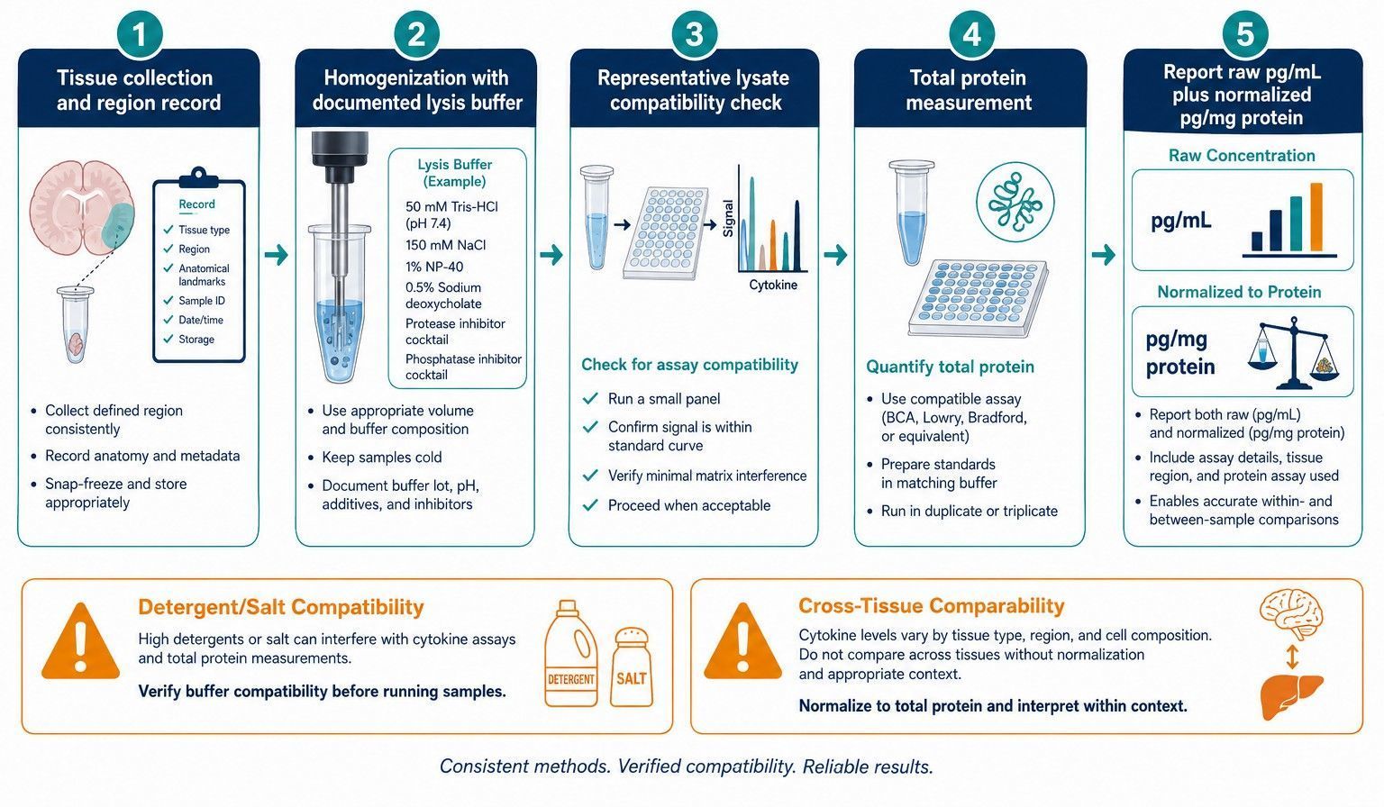 A tissue lysate cytokine study should lock collection, extraction, qualification, normalization, and reporting rules in sequence.
Total Protein Normalization for Tissue Cytokine Results
Total protein normalization relates each measured cytokine amount to the soluble protein recovered from the same lysate. It reduces variation caused by unequal extraction yield and variable dilution of the extract. It is especially useful when sample mass is limited or when the study compares matched tissue regions processed under one workflow.
The calculation should be transparent. If the cytokine concentration is reported by the assay in pg/mL and the corresponding extract contains mg/mL total protein, dividing the two produces pg/mg total protein. Preserve both inputs and the applied dilution factor in the report rather than supplying only a normalized endpoint.
What total protein normalization does and does not control
| Issue | How total protein normalization helps | What still needs study metadata |
|---|---|---|
| Different extraction yields | Reduces apparent differences caused by more or less soluble protein in an extract. | Tissue input mass, lysis volume, extraction chemistry. |
| Different tissue sizes | Provides a common soluble-protein denominator. | Wet weight, anatomical region, cellular composition. |
| Fibrosis, lipid, necrosis, or edema | Can make extract-to-extract comparisons more transparent. | Histology, composition context, and limitations of the denominator. |
| Residual blood content | Does not reliably remove blood-derived cytokine contribution. | Perfusion or collection record and interpretation boundary. |
| Assay matrix interference | Does not correct antibody-binding interference. | Matrix qualification and dilution/recovery evidence. |
Studies of brain tissue multiplexing have shown that both tissue processing and normalization influence the final cytokine value (Hulse et al., 2004). Total protein is a strong default when the goal is to compare extracted tissue content, but it should not be presented as proof that two biologically dissimilar tissues contain equivalent cellular material.
Choosing pg/mL, pg/mg Protein, and Wet Weight Reporting Units
No single reporting unit is universally correct. The appropriate unit follows the analytical and biological claim. A defensible data package may include more than one view: raw measured concentration for traceability, normalized concentration for comparison, and wet-weight or tissue-input metadata for context.
| Reporting approach | Best suited to | Main limitation |
|---|---|---|
| pg/mL extract | Assay traceability, dilution planning, and within-extract concentration. | Strongly dependent on extraction volume and yield. |
| pg/mg total protein | Comparing matched lysates where extraction yield differs. | Does not fully correct composition differences across tissues. |
| pg/mg wet tissue | Mass-based yield questions with rigorously standardized tissue input. | Can be distorted by water, lipid, blood, fibrosis, or extracellular matrix. |
| pg per defined tissue region | Microdissected or anatomically fixed samples. | Requires highly consistent sampling geometry and region definition. |
If the primary question concerns relative cytokine content in comparable lysates, choose pg/mg total protein as the main comparative unit and retain pg/mL in the analytical appendix. If the primary question is tissue-level yield from standardized, weighed samples, include wet-weight context as a secondary descriptor. Do not merge data reported under different denominators into one comparison without an explicit conversion rationale.
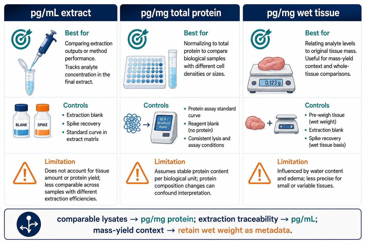 The reporting unit should be selected for the biological comparison, while raw extract concentration and normalization inputs remain traceable.
Making the Total-Protein Denominator Comparable
A total-protein denominator is only as comparable as the protein measurement behind it. Use one protein-assay method, one calibration approach, and one dilution rule within each direct comparison. The protein assay must also tolerate the intended lysis chemistry. A detergent or reducing agent that is acceptable for extraction can bias some protein-assay readouts, which would transfer directly into every normalized cytokine result.
Measure total protein from the same clarified lysate used for cytokine analysis whenever possible. If an aliquot is diluted for multiplex measurement, retain the aliquot identity and dilution factor so the cytokine and protein values can be reconciled. Do not normalize cytokine results to a protein value obtained from a separately processed piece of tissue unless the relationship is explicitly validated for the sampling design.
Predefine how values outside the useful measurement range will be treated. A cytokine concentration below the assay's reporting range should not become a precise normalized number merely because a protein denominator is available. Retain the original result flag, the dilution used, the protein value, and the final reported-unit rule. This creates a data package that can be rechecked if the cohort later needs a new comparison or a focused follow-up panel.
For a study with limited material, a sensible manifest links each sample ID to tissue region, lysate volume, total protein concentration, multiplex dilution, raw analyte concentration, normalized analyte value, and any qualifier. That structure is more useful for downstream interpretation than an isolated spreadsheet column of pg/mg values.
How to Make Tissue Cytokine Comparisons Interpretable
Comparable tissue cytokine data begin before assay submission. Hold constant the species, tissue region, collection window, pre-extraction storage, homogenization approach, buffer formula, and protein assay method within each direct comparison. Record deviations as data rather than relying on memory during interpretation.
For a multi-organ study, a separate qualification or stratified analysis may be preferable to one pooled conclusion. For example, adipose-rich, fibrotic, and highly vascular tissues can differ substantially in extraction behavior. If the scientific question is within-organ treatment response, analyze the organs in their own matched strata. If the question is cross-organ distribution, report that cross-organ normalization has biological limits.
Use the body-fluid and complex-matrix cytokine assay service as an entry point when the project includes tissue extracts alongside other challenging matrices. This allows the feasibility discussion to be matched to actual sample composition rather than to a generic tissue category.
Common Tissue Lysate Cytokine Profiling Mistakes
- Reporting pg/mL as though it is independent of extraction volume and protein yield.
- Using one lysis buffer across tissues without confirming that key analytes remain measurable and comparable.
- Normalizing after reviewing group differences rather than predefining the denominator.
- Comparing tissue types under one denominator without documenting composition and collection differences.
- Omitting buffer additives, total-protein values, tissue input, or dilution from the sample manifest.
- Assuming total protein normalization corrects matrix interference or blood contamination.
Planning a Tissue Lysate Cytokine Profiling Project
A project discussion should start with the biological comparison, not the panel size. Provide the tissue type and region, species, expected sample number, available mass or lysate volume, lysis-buffer composition, key cytokines, and intended primary unit. From there, the study can determine whether one multiplex panel is appropriate, whether feasibility data are needed, and which results should be delivered as raw concentrations, normalized values, and QC annotations.
Creative Proteomics can support multiplex cytokine profiling with study-specific review of tissue lysate compatibility, analyte selection, normalization logic, and report structure. The goal is a dataset whose comparison unit is aligned with the biological question rather than chosen after results are available.
Tissue Lysate Cytokine Study Design Checklist
Before submitting the full cohort, document the decisions that determine comparability: the exact tissue region and collection interval, whether blood removal or perfusion is part of the protocol, input mass or available lysate volume, complete lysis-buffer formulation, clarified-lysate handling, total-protein assay, proposed multiplex dilution, primary reporting unit, and intended comparison groups. These are not administrative details; each one sets a boundary on how the final cytokine difference can be interpreted.
Use the feasibility set to make a go/no-go decision on the final workflow. A practical go decision requires adequate detectability for the decision-critical analytes, acceptable behavior at the planned dilution, a protein denominator obtained consistently, and a reporting rule set before group labels are evaluated. If one of these conditions is not met, resolve it through buffer adjustment, sample dilution, a focused panel, or a narrower biological comparison. This approach protects both scarce tissue material and the value of the final cohort.
Frequently Asked Questions
Should tissue cytokine results be reported as pg/mL or pg/mg protein?
Use pg/mL to document the measured extract concentration, but use pg/mg total protein when the scientific question compares cytokine content across tissue lysates with potentially different extraction yields. Keep the original pg/mL values and the normalization denominator in the data package.
Can I use RIPA buffer for multiplex cytokine profiling?
Do not assume that a detergent-rich lysis buffer is compatible with a multiplex immunoassay. Its salts, detergents, and other additives can alter recovery or background, so confirm compatibility in a small matrix qualification exercise before scaling the study.
Is wet tissue weight an acceptable normalization denominator?
Wet weight can be informative for anatomical yield, but it does not correct for differences in extraction efficiency, edema, blood content, or composition. It is best treated as complementary metadata rather than an interchangeable substitute for total protein normalization.
Can cytokine data from different tissues be directly compared?
Only with a stated biological rationale and matched collection, extraction, normalization, and reporting rules. A pg/mg protein value does not by itself make a lymphoid tissue, adipose tissue, and fibrotic tissue biologically equivalent.
When should a tissue lysate project include matrix qualification?
Include it when the matrix is nonstandard, the lysis chemistry differs from the assay’s intended sample type, decision-critical analytes are near the lower range, or the study will compare several tissue types. A pilot should assess recovery and dilution behavior in representative extracts.
What information should be supplied with tissue lysate samples?
Provide tissue identity, species, collection interval, wet weight or input amount, lysis buffer and additives, total protein concentration, dilution, clarifying steps, storage history, and the intended reporting unit. These details determine what comparisons can be supported.
References:
- Stavnsbjerg C, et al. Matrix effect in tumor lysates – Does it affect your cytokine ELISA and multiplex analyses? Journal of Immunological Methods. 2022;500:113177.
- Hulse RE, et al. Optimization of multiplexed bead-based cytokine immunoassays for rat serum and brain tissue. Journal of Neuroscience Methods. 2004;136:87–98.
- Rosenberg-Hasson Y, et al. Effects of serum and plasma matrices on multiplex immunoassays. Immunologic Research. 2014;58:224–233.
- Ahnstedt H, et al. Sex differences in adipose tissue CD8+ T cells and regulatory T cells. Frontiers in Immunology. 2018;9:659.
- Lee JW, et al. Fit-for-purpose method development and validation for successful biomarker measurement. Pharmaceutical Research. 2006;23:312–328.