Introduction
Granulocyte macrophage colony stimulating factor (GM-CSF), also called CSF-2, is mainly derived from T cells, monocytes, macrophages, endothelial cells, and fibroblasts. Among them, human GM-CSF is a 22 kD glycoprotein consisting of 127 amino acids. GM-CSF mainly acts on myeloid progenitor cells, exerts biological effects by binding with its high-affinity receptors. It rapidly enters the cell cycle to differentiate into granulocytes and macrophages, and finally becomes mature cells, which can prolong the life of mature cells and enhance the function of mature cells. It can not only promote the proliferation, differentiation and maturation of hematopoietic precursor cells, but also stimulate other cells to varying degrees, such as antigen-presenting cells (APC), fibroblasts, and keratinocytes.
Mechanism and Function
GM-CSF is a polypeptide factor with a broad spectrum of effects, which has a proliferative effect on early hematopoietic progenitor cells. GM-CSF receptor(GM R) is a transmembrane glycoprotein, a member of the cytokine receptor superfamily. It consists of two subunits: α (GM Rα) and β (GM Rβ). They can be divided into extramembrane region, transmembrane region and cytoplasmic region. Among them, the α-subunit has ligand specificity, and can specifically bind to GM-CSF with a low affinity, but cannot transmit signals. Although the β subunit itself cannot bind to GM-CSF, it can form a receptor complex that binds to the ligand with high affinity with the α subunit and transmits the stimulation signal stimulated by the ligand, causing various biological behaviors of the cell. Because neither the α nor β subunits have tyrosine kinase domains, after the α subunits bind to the ligand, they must form a heterodimer with the β subunits through spatial allometry before they can stimulate the cells. Matrix tyrosine kinase is phosphorylated to transmit signals, so receptor dimerization is key to signal transduction.
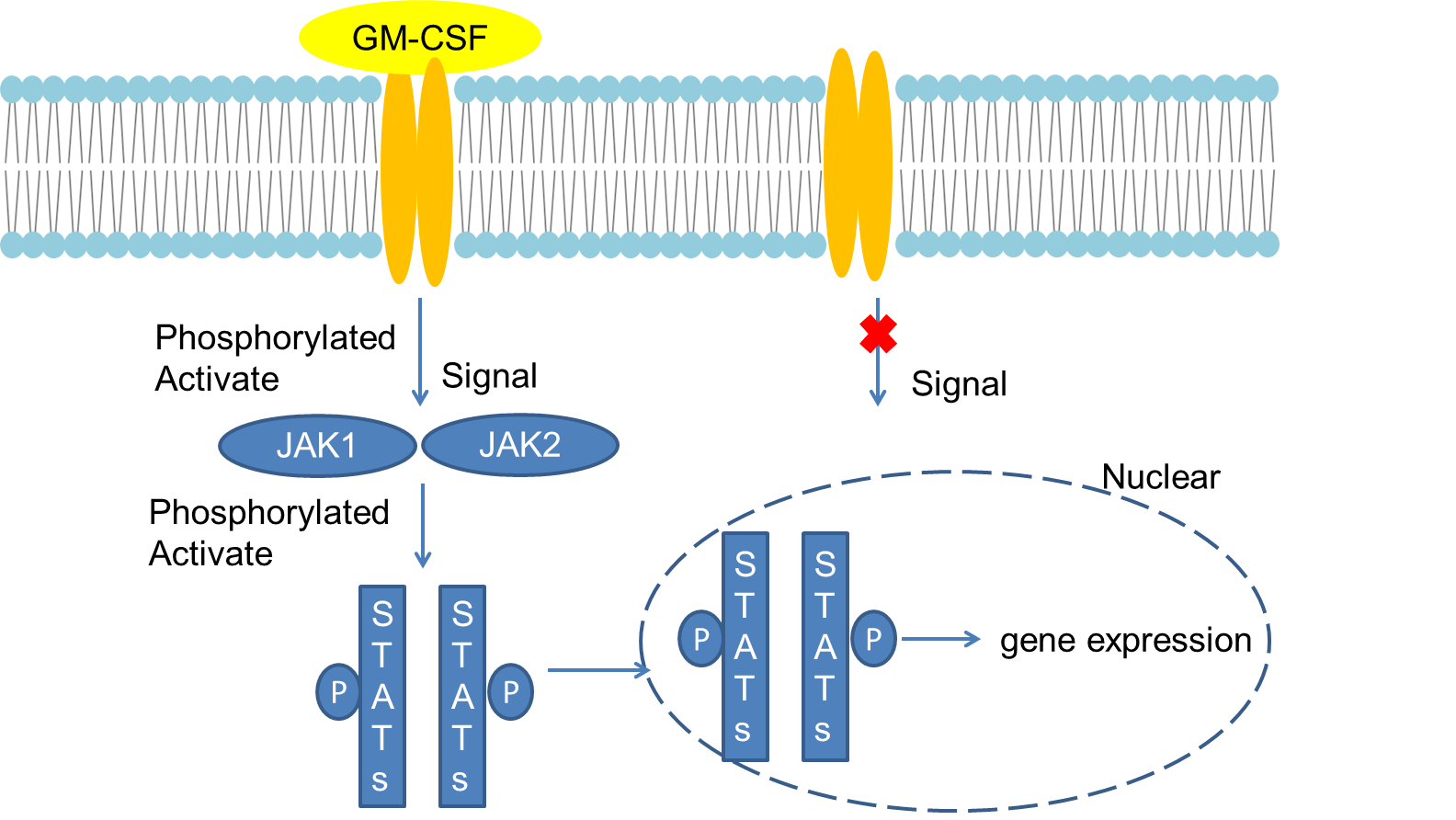
When it binds to the receptor, the receptor forms a heterodimer through conformational changes. After binding to the receptor, JAK activates receptor-JAK tyrosine phosphorylation. The SH2 domain of STATs then binds to the phosphorylated tyrosine site of the receptor complex. Under the action of JAKs, tyrosine at a specific site in STATs is phosphorylated and activated to form a dimer, enter the nucleus, bind to the promoter of the corresponding gene, and start gene expression.
GM-CSF mainly stimulates the growth and differentiation of bone marrow precursor cells, and stimulates bone marrow precursor cells to differentiate into granulocytes and monocytes. GM-CSF also has the function of lowering serum cholesterol, which can be used to treat myeloid cell proliferation syndrome. GM-CSF plays a key role in the development and maturation of dendritic cells and the proliferation and activation of T cells, linking innate and adaptive immune responses. In mice, treating melanoma cells expressing GM-CSF can increase the number of eosinophils, monocytes, macrophages, and lymphocytes, leading to a sustained antitumor response. GM-CSF facilitates the expansion of the DC1 population and increases the DC-mediated response to tumor cells. In vitro studies of human myeloid leukemia cells, in addition to promoting antigen presentation, GM-CSF also directs cells to the DC phenotype. GM-CSF is used to improve neutropenia in elderly patients with acute myeloid leukemia after induction chemotherapy.
 Fig 1. Mechanism of Signaling
Creative Proteomics can provide cytokine detection platform for scientific research. According to different purposes, our dedicated analysts will customize exclusive solutions for you. We aim to provide customers with high-quality and convenient services to help you accelerate the progress of your project.
Our cytokine detection service includes but is not limited to:
- One or more cytokines cytokines qualitative and quantitative detection
- Cytokines qualitative and quantitative detection of various species
- Cytokine antibodies qualitative and quantitative detection
Sample requirements
- Sample Types- Serum, plasma, etc.
- Sample Volume - It is optimal for 50 samples. This volume allows for triplicate testing of each sample.
Our advantages:
- Different detection methods can be selected based on different samples and requirements.
- Ensure the specificity and accuracy of the test by using high quality antibodies.
- Repeat the test to ensure the repeatability and accuracy of the experimental results.
- Feedback results are accurate and efficient
Technology platform:
We mainly provide the Luminex cytokine detection platform. Luminex uses fluorescently encoded microspheres with specific antibodies to different target molecules. The different microspheres can be combined freely to a certain extent so that up to 100 analytes can be tested multiple times simultaneously in a single experiment.
The Luminex cytokine assay platform has the following advantages:
- Multiple detection: simultaneous detection of 100 biological targets
- Short experiment time: 1-3 weeks
- High sensitivity: the lower limit of accurate quantification is as low as 0.1 pg/mL
- Save samples: only need a sample volume as low as 25 μL
- Time saving: the experiment process only takes 4 hours
For your different needs, we can also provide the following detection methods:
- Enzyme-linked immunosorbent assay (ELISA)
- Flow cytometry
Workflow

For more information about the GM-CSF detection service or need other detection requirements, please contact us.
References:
- Lilly M B, Zemskova M, et al. Distinct domains of the human granuIocyte-macrophage colony stimulating factor receptor α subunit mediate activation of Jak/Stat signaling and differentiation [J].Blood, 2001, 97:1662-1 670.
- Nosaka T, Kitamura T. Janus Kinases (JAKs) and Signal Transducers and Activations of Transcrption (STATs) in hematopoietic cells[J]. Int J Hematol, 2000, 71:309-319.

