
Introduction
Tumor necrosis factor-β (TNF-β), also known as lymphotoxin-α(LT-α), is a cytotoxic protein of the TNF superfamily, which is mainly expressed by T and B lymphocytes. TNF-β is encoded by the LTA gene in the human genome, and expression is highly inducible. The expressed TNF-β can either form a soluble homotrimeric molecule or form a heterotrimer with lymphotoxin-β (LT-β). By binding to LT-β, TNF-β can be anchored on the cell surface by the form LT-α 1 -β 2 complex. As a protein with multiple functions, TNF-β plays multiple roles in the innate immune system. According to existing studies, TNF-β has a strong antagonistic effect on cell proliferation ability and can destroy tumor growth, so it is an important cancer regulator. In addition, it is also involved in regulating physiological functions such as cell survival, differentiation, and apoptosis. When TNF-β is over-expressed, it can cause a long-term inflammatory response, which can lead to tumor development.
Mechanism and Function
As a member of the TNF family, TNF-β can bind to its related receptors to activate the NF-κB pathway, thereby achieving participation in immune regulation through the innate immune response. In order to activate TNF-β, TNF-β must form a LT-α 1 -β 2 complex with LT-β. This complex can bind the LT-β receptor and activate related signal transduction pathways, such as the NF-κB pathway. The signaling sequence of the NF-κB classic pathway is first activation of IKK complexes (IKKα, β, and γ), and then IKK complexes release the inhibition of RelA / p50 heterodimer by regulating IκBα phosphorylation and subsequent degradation. Finally, the RelA/ p50 heterodimer enters the nucleus and induces the expression of pro-inflammatory cytokines, chemokines and adhesion molecule-related genes. On the other hand, the alternative NF-κB path depends on NIK and IKKα, and p100 is processed into p52 with the participation of IKKα. Then p52 and RelB form a RelA / p52 heterodimer and enter the nucleus, thereby activating genes involved in the regulation of lymphoid organogenesis and homeostasis. In addition, other mechanisms of LTβR-induced cell death include the production of reactive oxygen species (ROS), ASK-1, and caspase-independent or caspase-dependent apoptosis. LTβR has also been shown to activate the JNK signaling pathway and induce AP-1 regulated gene expression and cell death.
Interestingly, TNF-β both inhibits tumor growth and promotes tumor development. Recent studies have shown that TNF-β signaling can promote inflammatory responses, but prolonged inflammatory responses can also cause severe cellular damage and increase the risk of some diseases, including cancer. Therefore, mutations in regulatory factors of TNF-β signaling pathway can cause disruption of the cell signaling regulatory network and lead to cancer cell production. These mutations usually occur on the LT-α1-β2 complex and the LT-β receptor, the downstream NF-κB alternative pathway is sustaining activated. Studies have further confirmed the existence of this mechanism. And after knocking out the LT-β receptor, it showed obvious effects of inhibiting tumor growth and reducing angiogenesis. Therefore, TNF-β and its downstream signaling pathway are involved in the process of cytokines affecting tumor development and metastasis, and this process will show dual effects on tumors according to various factors.
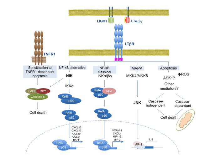 Figure 1. Mechanism of TNF-βSignaling Pathway.
Creative Proteomics can provide cytokine detection platform for scientific research. According to different purposes, our dedicated analysts will customize exclusive solutions for you. We aim to provide customers with high-quality and convenient services to help you accelerate the progress of your project.
Our cytokine detection service includes but is not limited to:
- Qualitative and quantitative analysis of single cytokines
- Qualitative and quantitative analysis of multiple cytokines
- Qualitative and quantitative detection of cytokines in various species
- Qualitative and quantitative detection of cytokine antibodies
Sample requirements
- Sample Types - Blood, serum, plasma, cell culture supernatant, cell lysate, cell culture medium, tissue homogenate, urine, tumor, etc.
- Sample Volume - It is optimal for at least 200µl of each sample. This volume allows for triplicate testing of each sample.
Our advantages:
- There are multiple detection methods that can be selected based on different samples and requirements.
- High-quality antibodies are used in the detection process to improve the specificity and accuracy of the detection.
- Repeat testing to ensure reproducibility and accuracy of test results.
- Professional and efficient feedback results.
Technology platform:
We mainly provide the Luminex cytokine detection platform. Luminex uses fluorescently encoded microspheres with specific antibodies to different target molecules. The different microspheres can be combined freely to a certain extent so that up to 100 analytes can be tested multiple times simultaneously in a single experiment.
The Luminex cytokine assay platform has the following advantages:
- Multiple detection: simultaneous detection of 100 biological targets
- Short experiment time: 1-3 weeks
- High sensitivity: the lower limit of accurate quantification is as low as 0.1 pg/mL
- Save samples: only need a sample volume as low as 25 μL
- Time saving: the experiment process only takes 4 hours
For your different needs, we can also provide the following detection methods:
- Enzyme-linked immunosorbent assay (ELISA)
- Flow cytometry
Workflow

For more information on TNF-β testing services or to require additional testing requirements, please contact us.
References:
- Bauer J, et al. Lymphotoxin, NF-ĸB, and cancer: the dark side of cytokines. Digestive Diseases. 2012, 30 (5): 453–68.
- Müller JR, Siebenlist U. Lymphotoxin beta receptor induces sequential activation of distinct NF-kappa B factors via separate signaling pathways. The Journal of Biological Chemistry. 2003, 278 (14): 12006–12.
- Mónica T. Fernandes, et al. Context-dependent roles for lymphotoxin-β receptor signaling in cancer development. Biochimica et Biophysica Acta. 2016, 1865: 204–219.