
Introduction
Interleukin-1 (IL-1) was first described as a protein that causes fever, called human leukocyte pyrogens. It is also known as lymphocyte activating factor. IL-1 has two agonists, IL-1α and IL-1β, and a receptor antagonist IL-1Ra. IL-1 is mainly produced by monocytes, macrophages in the blood, and various epithelial cells. Besides, endothelial cells and mesenchymal cells can also produce IL-1. IL-1 has the function of regulating immune response, inflammatory response and hematopoiesis. It is induced by various stimulating factors (including antigen, endotoxin, bacteria and viruses), plays an important role in the pathogenesis of acute and chronic inflammation, and is closely related to the pathological processes of various diseases. IL-1 is also involved in the body's hematopoietic system, the response of the nervous and endocrine systems, and certain anti-tumor pathophysiological processes. In summary, monitoring IL-1 can help to understand the body's immune regulation ability, and can provide a reliable basis for disease diagnosis, therapeutic observation and prognosis.
Mechanism and Function
IL-1 is a pleiotropic cytokine that primarily affects inflammation and immune response. It also regulates other physiological functions of the body and has an important influence on the pathogenesis of the disease. The IL-1α and IL-1β sequences are not highly homologous, but they bind to the same receptor complex and have similar biological activities. In fact, there are still some differences in specific mechanisms of action and biological functions. IL-1α is mainly cell-bound and active, including intracellular precursors (pro IL-1α) and membrane-bound IL-1α, with few secreted forms. IL-1α precursor is produced under normal physiological conditions and stored in the cytoplasm and on the cell membrane, and the expression level is elevated when inflammation occurs. IL-1β is only secreted and active, its precursor is inactive, and there is no membrane-bound type. IL-1β precursor is not produced under normal physiological conditions, and it is secreted only when stimulated. Besides, its physiological action is exerted after cleaving by caspase-1.
IL-1α and IL-1β bind to the same receptor IL-1RI (IL-1RI is a signaling receptor). After IL-1 binds to IL-1RI, another protein, the IL-1R accessory protein (IL-1RAcP), is recruited to form a complex with IL-1/IL-1RI. And hybrid dimers of IL-1RI and IL-1RAcP lead to signal transduction. The dimer first activates the IL-1 receptor-associated kinase (IRAK), and is mediated by nuclear transcription factors NF-κB, AP-1, JNK, p38 MAPK, hsp 27, etc., which ultimately leads to the activation of nuclear genes. IL-1Ra also binds to IL-1RI, it does not activate further signaling, thereby blocking IL-1 action.
IL-1 is secreted by macrophages and initiates an inflammatory response to induce expression of proinflammatory genes such as COX-2. And it has different enhancement effects on the proliferation, differentiation and function of different cells in the body, such as NK cells, macrophages, granulocytes, and special immunocompetent cells including T cells and B cells. IL-1, also known as hemopoietin-1, is a hematopoietic factor essential for maintaining hematopoietic function, which can enhance the invasiveness of existing tumors leading to tumor spread and metastasis.
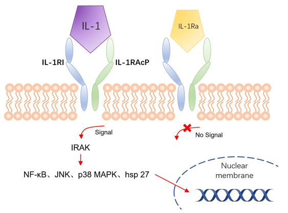 Fig 1. Mechanism of Signaling
Creative Proteomics can provide cytokine detection platform for scientific research. According to different purposes, our dedicated analysts will customize exclusive solutions for you. We aim to provide customers with high-quality and convenient services to help you accelerate the progress of your project.
Our cytokine detection service includes but is not limited to:
- Single cytokine qualitative and quantitative detection
- Multiple cytokines qualitative and quantitative detection
- Cytokines qualitative and quantitative detection of multiple species
- Cytokine antibodies qualitative and quantitative detection
Sample requirements
- Sample Types-Blood, serum, plasma, cell culture supernatant, cell lysate, cell culture medium, tissue homogenate, urine, tumor, etc.
- Sample Volume - It is optimal for at least 200µl of each sample. This volume allows for triplicate testing of each sample.
Our advantages:
- A variety of detection methods are available, which can be selected according to different samples and requirements.
- High-quality antibodies are used in the detection process to improve detection specificity and accuracy.
- Repeat testing to ensure repeatability and accuracy of experimental results.
- The feedback results are professional and efficient.
Technology platform:
We mainly provide the Luminex cytokine detection platform. Luminex uses fluorescently encoded microspheres with specific antibodies to different target molecules. The different microspheres can be combined freely to a certain extent so that up to 100 analytes can be tested multiple times simultaneously in a single experiment.
The Luminex cytokine assay platform has the following advantages:
- Multiple detection: simultaneous detection of 100 biological targets
- Short experiment time: 1-3 weeks
- High sensitivity: the lower limit of accurate quantification is as low as 0.1 pg/mL
- Save samples: only need a sample volume as low as 25 μL
- Time saving: the experiment process only takes 4 hours
For your different needs, we can also provide the following detection methods:
- Enzyme-linked immunosorbent assay (ELISA)
- Flow cytometry
Workflow
For more information about the IL-1 detection service or need other detection requirements, please contact us.
References:
- Murray K N, et al. Interleukin-1 and acute brain injury. Frontiers in Cellular Neuroscience, 2015, 9.
- Rivers-Auty J, et al. Redefining the ancestral origins of the interleukin-1 superfamily. Nature Communications, 2018, 9(1):1156.