
Introduction
Erythropoietin (EPO), also known as red blood cell stimulating factor, is a human-derived glycoprotein hormone that stimulates red blood cell production. Normal human body has a certain amount of erythropoietin, which is mainly produced by the kidney and a small amount is produced by the liver. According to the source, it can be divided into endogenous and exogenous erythropoietin. Natural erythropoietin molecules include a polypeptide portion and a sugar chain portion. Erythropoietin, which is present in plasma, consists of 165 amino acids. Different types of erythropoietin amino acid polypeptides are consistent, with a molecular weight of 18235Da. They are connected by disulfide bonds to form four stable α helix structures, which is the spatial conformation necessary to maintain the biological activity of erythropoietin. Hypoxia can stimulate the production of erythropoietin. Recombinant human erythropoietin is used to treat anemia associated with renal insufficiency, acquired immune deficiency syndrome / AIDS itself or anemia caused by treatment, anemia associated with malignant tumors, and rheumatism Anemia and other anemia.
Mechanism and Function
Erythropoietin is a colony-stimulating factor and belongs to the type I cytokine superfamily. The human EPO gene is located in the 22 region of the long arm of chromosome 7, and contains 5 exons and 4 introns, encoding a protein of 193 amino acids. This protein is a precursor of active EPO, and its biological effects can only be exerted by cutting off 27 amino acids and removing terminal amino acids. The presence of sugar chains and sialic acid is a necessary condition for EPO to exert its biological activity in the body. In the fetus, EPO is mainly synthesized by the liver. After birth, EPO is mainly secreted and synthesized by renal interstitial fibroblasts. Adults synthesize about 90% of total EPO in the kidney, and the liver and central nervous system synthesize the remaining 10%. EPO is synthesized by astrocytes and neurons in the central nervous system, and EPO is synthesized by fat-storaging cells (Ito) and liver cells in the liver. The physiological function of EPO is mainly combined with the surface receptors (called EPOR) of Colony Forming Unit-erythroid, which promote the differentiation of erythroid directional stem cells into erythroblasts, hemoglobin synthesis of nucleated red blood cells, and release of red blood cells.
EPOR is a transmembrane protein composed of 484 amino acids with a relative molecular mass of 55,000. Its gene is located in the short arm of chromosome 19, belongs to the cytokine receptor superfamily, and is a dimeric type I receptor. An EPO molecule binds to the EPOR homodimer through two different sites, thereby activating EPOR, causing the activation of tyrosine kinase 2 (JAK2) in the cytoplasm, and then inducing signaling pathways including the transcriptional activation factor (STAT). Finally, it can inhibit the apoptosis of Colony Forming Unit-erythroid and promote their proliferation and differentiation.
EPO can be used to treat anemia associated with renal insufficiency, anemia caused by acquired immunodeficiency syndrome itself or anemia caused by treatment, anemia associated with malignant tumors, anemia caused by rheumatism, and rheumatoid arthritis and severe. In addition, EPO can also be used to prevent anemia. Besides, for patients with chronic renal failure associated with anemia, including patients with and without dialysis, EPO can increase or maintain red blood cell levels and reduce blood transfusion. Although EPO can avoid dependence on blood transfusion, it cannot replace emergency blood transfusion. EPO can be used to improve the symptoms of anemia in patients with globin production anemia (thalassemia) or precocious anaemia, can accelerate the recovery of hematopoietic function after bone marrow transplantation, and can also be used for anemia caused by aluminum overload.
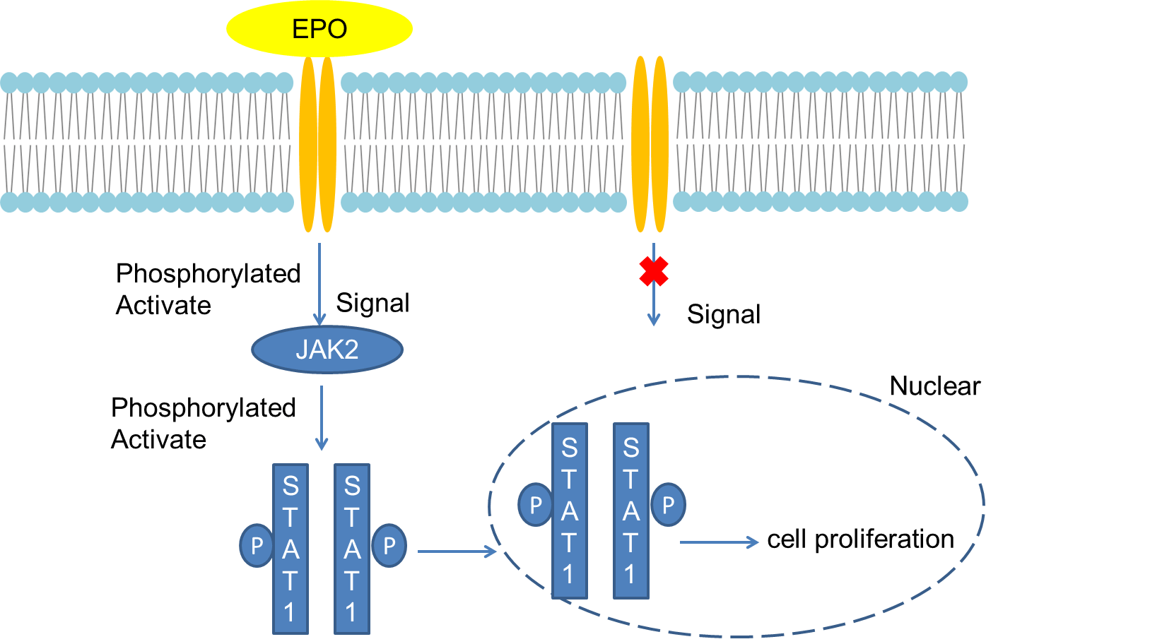 Fig 1. Mechanism of Signaling
Creative Proteomics can provide cytokine detection platform for scientific research. According to different purposes, our dedicated analysts will customize exclusive solutions for you. We aim to provide customers with high-quality and convenient services to help you accelerate the progress of your project.
Our cytokine detection service includes but is not limited to:
- One or more cytokines cytokines qualitative and quantitative detection
- Cytokines qualitative and quantitative detection of various species
- Cytokine antibodies qualitative and quantitative detection
Sample requirements
- Sample Types-Blood, serum, plasma, cell culture medium, tissue homogenate, etc.
- Sample Volume - It is optimal for 50 samples. This volume allows for triplicate testing of each sample.
Our advantages:
- Different detection methods can be selected based on different samples and requirements.
- Ensure the specificity and accuracy of the test by using high quality antibodies.
- Repeat the test to ensure the repeatability and accuracy of the experimental results.
- Feedback results are accurate and efficient
Technology platform:
We mainly provide the Luminex cytokine detection platform. Luminex uses fluorescently encoded microspheres with specific antibodies to different target molecules. The different microspheres can be combined freely to a certain extent so that up to 100 analytes can be tested multiple times simultaneously in a single experiment.
The Luminex cytokine assay platform has the following advantages:
- Multiple detection: simultaneous detection of 100 biological targets
- Short experiment time: 1-3 weeks
- High sensitivity: the lower limit of accurate quantification is as low as 0.1 pg/mL
- Save samples: only need a sample volume as low as 25 μL
- Time saving: the experiment process only takes 4 hours
For your different needs, we can also provide the following detection methods:
- Enzyme-linked immunosorbent assay (ELISA)
- Flow cytometry
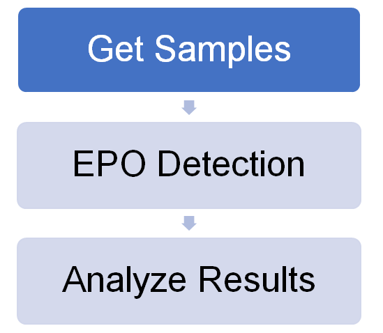
Workflow
For more information about the EPO detection service or need other detection requirements, please contact us.
References:
- Cokie VP, Bhattacharya B, et al. JAK-STAT and AKT pathway-coupled genes in crythroid progenitor cells through ontogeny. J Transl Med, 2012,10:11
- Jelkmann W, Bohlius J, et al. The crythropoietin receptor in normal and cancer tissues. Crit Rev Oncol Hematol, 2008, 67(1):39-61.