
Introduction
Interleukin-9 is released by helper T cells and can also be produced by T cells transformed by T cell leukemia virus 1 (HTLV-1), which is a type of T cell growth factor. It is expressed in human eosinophils, natural killer cells, Th9 cells, Th17 cells and Treg cells. In addition to stimulating the growth of T cells, it can also support the growth of hematopoietic progenitor cells, expand the response of bone marrow mast cells to IL-3, and promote various functions such as the growth of certain leukemia cell lines, thymoma cell lines, and T cells. IL-9 can stimulate airway epithelial cells (AEC) to secrete certain proteins and activate the expression of AEC mucin gene subsets, causing AEC to secrete excessive mucusIL-9 can also directly act on AEC to produce chemokines and promote the accumulation of inflammatory cells in the lungs, which is related to human asthma.
Mechanism and Function
IL-9 acts through a receptor on the surface of the target cell membrane. The receptor for IL-9 (IL-9R) exists on the surface of a variety of cell membranes. It is a heterodimer structure composed of a specific α-subunit ligand and A common gamma chain consists of two strands. IL-9 can promote tumor cell proliferation by activating the STAT3 phosphorylation level and then activating the STAT3 signaling pathway. The activation of IL-9 promotes the phosphorylation of protein tyrosine kinase 3 (JAK3), thereby causing signal transduction in downstream regions and activation of transcription activation factor (STAT), especially STAT1 homodimer, STAT5 homodimer and STAT1-STAT3 heterodimer. STAT activation is necessary in IL-9-induced T cell proliferation.
Under specific circumstances, IL-9 can enhance the suppressive function of Treg cells. The mechanism of IL-9 acting on Treg is through the activation of STAT3 and STAT5 signals. A lot of evidence shows that STAT3 and STAT5 signaling pathways play a key regulatory role in the development of Treg. Therefore, IL-9 can affect the immune response and Th17 / Treg balance.
IL-9 is also related to allergic asthma. IL-9 participates in the development of the disease by promoting the proliferation of mast cells and the production of IL-13, and subsequently increasing the release of mucus and participating in airway hyperresponsiveness. And IL-9 does not rely on inflammatory responses to directly induce these changes. IL-9 plays a role in peribronchial fibrosis, and overexpression of IL-9 can aggravate fibrosis around the bronchi. In addition, it can directly act on red blood cell-oriented liver cells, and has the effect of selectively stimulating red blood cell production.
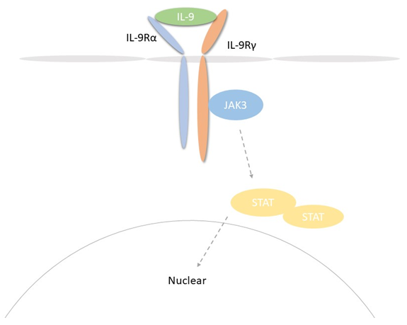 Fig 1. Mechanism of Signaling
Creative Proteomics can provide cytokine detection platform for scientific research. According to different purposes, our dedicated analysts will customize exclusive solutions for you. We aim to provide customers with high-quality and convenient services to help you accelerate the progress of your project.
Our cytokine detection service includes but is not limited to:
- Quantitative and qualitative detection of cytokines in different species
- Quantitative and qualitative detection of cytokine antibodies
- Quantitative and qualitative detection of single/mulltiple cytokines
Sample requirements
- Sample Types-Blood, serum, plasma, cerebrospinal fluid, cell culture supernatant, tissue homogenate, cell culture medium, urine, tumor, etc.
- Sample Volume - It is optimal for at least 200µl of each sample. This volume allows for triplicate testing of each sample.
Our advantages:
- Efficient design: Multiplex reaction detecting in various methods are available, which can be selected according to different samples and requirements.
- Sensitive detection: High-quality antibodies are used in the detection process to improve detection specificity and accuracy.
- Reliable results: The feedback results are professional and efficient.
Technology platform:
We mainly provide the Luminex cytokine detection platform. Luminex uses fluorescently encoded microspheres with specific antibodies to different target molecules. The different microspheres can be combined freely to a certain extent so that up to 100 analytes can be tested multiple times simultaneously in a single experiment.
The Luminex cytokine assay platform has the following advantages:
- Multiple detection: simultaneous detection of 100 biological targets
- Short experiment time: 1-3 weeks
- High sensitivity: the lower limit of accurate quantification is as low as 0.1 pg/mL
- Save samples: only need a sample volume as low as 25 μL
- Time saving: the experiment process only takes 4 hours
For your different needs, we can also provide the following detection methods:
- Enzyme-linked immunosorbent assay (ELISA)
- Flow cytometry
Workflow
For more information about the IL-9 detection service or need other detection requirements, please contact us.
References:
- Temann U A , Ray P , Flavell R A . Pulmonary overexpression of IL-9 induces Th2 cytokine expression, leading to immune pathology. Journal of Clinical Investigation, 2002, 109(1):29-39.
- Nowak E C , Weaver C T , et al. IL-9 as a mediator of Th17-driven inflammatory disease. Journal of Experimental Medicine, 2009, 206(8):1653-1660.