
Introduction
Chemokines are a class of factors or signaling proteins produced by some cells and secreted outside the cell. They are called chemokines because they induce directed movement of cells with corresponding receptors on the surface. Previous studies have shown that chemokines have four cysteine residues in their conserved positions. These residues are very important for the formation of the spatial structure of chemokines. Therefore, according to the different distribution of cysteine residues, scientists divided chemokines into four main subfamilies: CXC, CC, CX3C, and XC. Among them, the structure of the CX3C chemokine is that there are three amino acids between two cysteine, which is called CX3C chemokine (or d-chemokine). The only CX3C chemokine discovered to date is called fractalkine (or CX3CL1). It can both secrete and bind to the surface of cells that express it, so it is both a chemotactic agent and an adhesion molecule.
Mechanism and Function
It was found that the chemokine receptor is a seven-transmembrane G protein-coupled receptor that mediates the biological activity of chemokines. Most of these receptors exhibit promiscuous binding properties, that is, several different chemokines send signals through the same receptor. So far, CX3C chemokines have a CX3C receptor, CX3CR1, which is the receptor for fractalkine. It is a protein encoded by the CX3CR1 gene in humans. As the name suggests, this receptor binds to the chemokine CX3CL1 (also known as neurokinin or fractalkine). Through in-depth research on it, scientists found that its downstream signaling mechanism is similar to other subgroups, and it is through the binding of chemokines to chemokine receptors on the surface of target cells, which then activates the intracellular coupling of G protein to cause G Protein subunits are released. Free G protein subunits activate phospholipase C (PLC). The activated phospholipase C cleaves phosphatidylinositol (4,5) -diphosphate (PIP2) into inositol triphosphate (IP3) and diacylglycerol ( DAG). Among them, IP3 can cause changes in intracellular calcium ions and subsequent signal transduction, while DAG can activate protein kinase C (PKC). These events can promote subsequent cascades of related intracellular signals that ultimately cause the adhesion and migration of leukocytes.
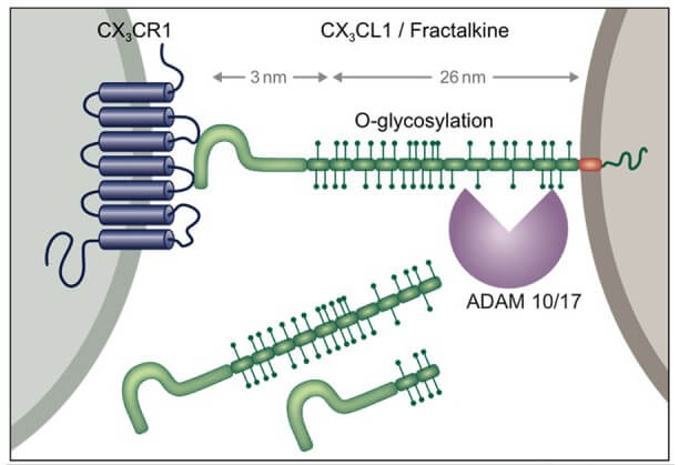 Figure 1. Schematic of CX3C chemokine family.
Creative Proteomics can provide cytokine detection platform for scientific research. According to different purposes, our dedicated analysts will customize exclusive solutions for you. We aim to provide customers with high-quality and convenient services to help you accelerate the progress of your project.
Our cytokine detection service includes but is not limited to:
- Qualitative and quantitative analysis of single cytokines
- Qualitative and quantitative analysis of multifarious cytokines
- Qualitative and quantitative detection of cytokines in different species
- Identification and quantitative determination of cell factor antibody
Sample requirements
- Sample Types - Blood, serum, plasma, cell culture supernatant, cell lysate, cell culture medium, tissue homogenate, urine, tumor, etc.
- Sample Volume - It is optimal for at least 200µl of each sample. This volume allows for triplicate testing of each sample.
Our advantages:
- Diversiform test methods can be selected according to samples and requirements.
- High-quality antibodies are used for test to ensure the specificity and accuracy of the results.
- Repeat test to ensure reproducibility and accuracy.
- Professional and efficient feedback results.
Technology platform:
We mainly provide the Luminex cytokine detection platform. Luminex uses fluorescently encoded microspheres with specific antibodies to different target molecules. The different microspheres can be combined freely to a certain extent so that up to 100 analytes can be tested multiple times simultaneously in a single experiment.
The Luminex cytokine assay platform has the following advantages:
- Multiple detection: simultaneous detection of 100 biological targets
- Short experiment time: 1-3 weeks
- High sensitivity: the lower limit of accurate quantification is as low as 0.1 pg/mL
- Save samples: only need a sample volume as low as 25 μL
- Time saving: the experiment process only takes 4 hours
For your different needs, we can also provide the following detection methods:
- Enzyme-linked immunosorbent assay (ELISA)
- Flow cytometry
Workflow
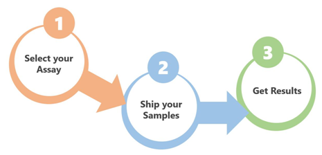 Fig 2. workflow
For more information on chemokines CX3C subfamily detection service or to require additional testing requirements, please contact us.
References:
- KYochai Wolf, Simon Yona, et al. Microglia, seen from the CX3CR1 angle. Frontiers in Cellular Neuroscience. 2013, doi: 10.3389/fncel.2013.00026
- Ono SJ, Nakamura T, Miyazaki D. Chemokines: roles in leukocyte development, trafficking, and effector function. The Journal of Allergy and Clinical Immunology. 2003.