
Introduction
Interleukin 36 (IL-36) belongs to IL-1 family and is an important mediator of inflammatory diseases. IL-36 includes IL-36α, IL-36β, IL-36γ and IL-36 receptor antagonists (IL-36Ra). IL-36Ra, IL-36α, IL-36β and IL-36γ were named as members of IL-1 family 5, 6, 8 and 9, respectively, and they all mediate signal transmission by IL-1 receptor-related protein (IL-1Rrp2) and IL- 1 receptor accessory protein (IL-1RAcP). IL-36Ra can prevent the activation of IL-36 receptor signal, so it has an inhibitory effect on the immune response of inflammatory diseases. IL-36 is involved in the occurrence and development of various inflammatory diseases, such as psoriasis, localized hair loss, joint inflammation, lung inflammation, and other inflammatory diseases.
Mechanism and Function
IL-36 belongs to the interleukin 1 family, which is a group of structurally related and functionally related cytokines, mainly related to the occurrence and development of inflammatory diseases such as skin inflammation. IL-36 family cytokines are only expressed in a few tissues and cells, including keratinocytes, bronchial epithelial cells, brain tissues and monocytes / macrophages. IL-36α, IL-36β and IL-36γ are high expression in human skin and rats esophagus, but low expression in mouse toes. IL-36α and IL-36β are expressed in T lymphocytes, and IL-17 and tumor necrosis factor (TNF) can induce IL-36α, IL-36β and IL-36γ expression in keratinocytes, which can be enhance by IL-22. Moreover, epidermal growth factor can regulate the expression of IL-36α and IL-36β in the skin. IL-36β can induce its own expression, because it contains autocrine and paracrine cycles. IL-36Ra is expressed in almost all keratinocytes. IL-36α is expressed in trachea, thymus, fetal colon, spleen tissue and mouse stomach tissue. IL-36β is expressed in human fetal colon and skeletal muscle. IL-36γ is lowly expressed in human uterus and tracheal tissues, but highly expressed in rat tissues and cells.
The mechanism of IL-36Ra antagonism is very similar to IL-1Ra. IL-36Ra first binds IL-1Rrp2, and then hinders the recruitment of IL-1RAcP and the formation of a functional signaling complex, thereby exerting its antagonistic effect. The activity of IL-36Ra depends on the excision of its N-terminal methionine. IL-36Ra has complete antagonist activity starting from Val-2. It can not only inhibit IL-36γ but also inhibit IL-36α and IL-36β, and its Val-2 is located at the N-terminal of all IL-1F members, a common A-X-Asp conservatively repeated 9th amino acid. The excision of IL-36α, IL-36β and IL-36γ N-terminal methionine can increase their activity by 1000-10000 times. The amount of IL-36Ra required for excised IL-36γ is 100-1000 times greater. Furthermore, the extracellular domain of IL-1Rrp2 is necessary for IL-36Ra to bind and exert its antagonistic effect.
IL-36 has a significant effect on dendritic cells and T cells. In mouse dendritic cells, IL-36R agonists can up-regulate CD80, CD86 and MHCⅡ, thereby inducing the production of IL-6 and IL-12, and the production of IL-6 and IL-12 mainly depends on IL-36R Signal pathway. IL-36α, IL-36β and IL-36γ activate mitogen-activated protein kinases (MAPK pathway) through IL-1Rrp2 and IL-1RAcP to produce a large number of cytokines, chemokines, adhesion molecules and enzymes and other inflammatory mediators, which in turn mediate Inflammation.
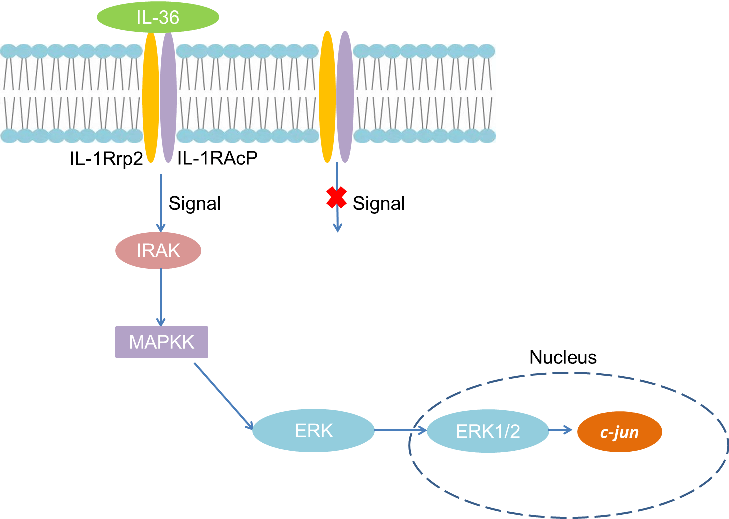 Fig 1. Mechanism of Signaling
Creative Proteomics can provide cytokine detection platform for scientific research. According to different purposes, our dedicated analysts will customize exclusive solutions for you. We aim to provide customers with high-quality and convenient services to help you accelerate the progress of your project.
Our cytokine detection service includes but is not limited to:
- One or more cytokines cytokines qualitative and quantitative detection
- Cytokines qualitative and quantitative detection of various species
- Cytokine antibodies qualitative and quantitative detection
Sample requirements
- Sample Types-Blood, serum, plasma, cell culture medium, tissue homogenate, etc.
- Sample Volume - It is optimal for 50 samples. This volume allows for triplicate testing of each sample.
Our advantages:
- Different detection methods can be selected based on different samples and requirements.
- Ensure the specificity and accuracy of the test by using high quality antibodies.
- Repeat the test to ensure the repeatability and accuracy of the experimental results.
- Feedback results are accurate and efficient
Technology platform:
We mainly provide the Luminex cytokine detection platform. Luminex uses fluorescently encoded microspheres with specific antibodies to different target molecules. The different microspheres can be combined freely to a certain extent so that up to 100 analytes can be tested multiple times simultaneously in a single experiment.
The Luminex cytokine assay platform has the following advantages:
- Multiple detection: simultaneous detection of 100 biological targets
- Short experiment time: 1-3 weeks
- High sensitivity: the lower limit of accurate quantification is as low as 0.1 pg/mL
- Save samples: only need a sample volume as low as 25 μL
- Time saving: the experiment process only takes 4 hours
For your different needs, we can also provide the following detection methods:
- Enzyme-linked immunosorbent assay (ELISA)
- Flow cytometry
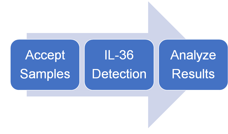
Workflow
For more information about the IL-36 detection service or need other detection requirements, please contact us.
References:
- Gresnigt MS, van de Veerdonk FL. Biology of IL-36 cytokines and their role in disease. Semin Immunol, 2013, 25(6):458-465.
- Marrakchi S,et al. Interleukin-36-receptor antagonist deficiency and generalized pustular psoriasis. N Engl J Med, 2011, 365(7):620-628.