
Tumor necrosis factor TNF cell signaling pathway
Based on Luminex technology platform, Creative Proteomics provides analysis services for key targets of TNF cell signaling pathway.
Tumor necrosis factor (TNF) is a cytokine produced naturally by macrophages in response to bacterial infection or other immune sources. Mainly produced by activated monocytes/macrophages, it can kill and inhibit tumor cells, promote neutrophil phagocytosis, fight infection, cause fever, induce acute protein synthesis in hepatocytes, and promote myeloid leukemia cells to differentiate into macrophages It promotes cell proliferation and differentiation, is an important inflammatory factor, and participates in the pathological damage of certain autoimmune diseases. Tumor necrosis factor (TNF) has a wide range of biological activities, and plays an important role in endotoxin shock, moxibustion, immune regulation, cell proliferation, cytotoxic activity and antiviral activity.
Tumor necrosis factor (TNF) is divided into two types according to its source and structure:
- TNF-α is mainly produced by macrophages. LPS is a strong stimulant that induces its production. T cells and NK cells can also secrete TNF-α under the action of certain stimulating factors (such as PMA); TNF-α is mainly produced by single Nuclear-secreted by macrophages.
- TNF-β is mainly produced by activated T cells. T cells can produce high levels of TNF-β under the stimulation of antigens and mitogens. TNF-β is mainly secreted by activated T lymphocytes, and both have similar pyrogenicity.
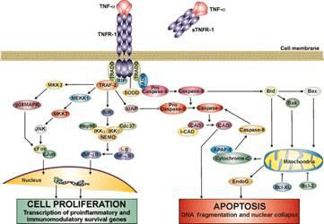
TNF first binds to the extracellular domain of TNF-R1 to cause trimerization of TNF-R1. Trimeric TNF-R1 recruits downstream signaling proteins such as TRADD, FADD, TRAFZ, and RIP through its aggregated intracellular death domain to assemble into a huge TNF-R1 signaling complex. This complex activates the downstream components. Signal transduction pathway, and finally activate the transcription factor NF-KB, JNK kinase, and induce cell apoptosis. Tumor necrosis factor (TNF), as a type of cytokine that can directly cause tumor cell death, works in concert with interferon to kill tumor cells.
Our detectable targets:
| JAK1 | MAPK | TYK2 | STAT1 | STAT2 | STAT3 |
| STAT5 | IRAK4 | JNK | MYD88 | Rac1 | TLR4 |
| CREB | IRF3 | p50 | NAP1 | Rel | TLR9 |
| FADD | IRF7 | Mda-5 | NFκB | p65 | TRAF3 |
| GAS | IRF9 | MEKK1 | p38 | RIP1 | Vav |
| IRAK1 | IRF5 | MEK3 | p38MAPK | PKR | TRAF6 |
| IPS-1 | IRS1 | MEK6 | PI3K | Tak1 | TRAM |
Technology platform
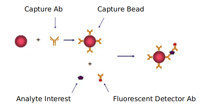
We provide Luminex technology for TNF cell signaling pathway analysis.
Luminex technology is a multifunctional liquid phase analysis platform developed on the basis of colored microspheres, laser technology, applied fluidics and high-speed digital signal processing technology. The core is to encode polypropylene microspheres or magnetic microspheres with fluorescent dyes. By adjusting the different ratios of the two fluorescent dyes, up to 100 microspheres with different fluorescence spectra can be obtained. Each kind of microspheres is covalently cross-linked. Capture antibodies against specific antigens.
TNF is a cytokine produced naturally by macrophages in response to bacterial infection or other immune sources. Tumor necrosis factor (TNF) cell signaling plays a role in promoting the phagocytosis of neutrophils, resisting infection, causing fever, inducing acute protein synthesis in hepatocytes, promoting the differentiation of myeloid leukemia cells into macrophages, and promoting cell proliferation and differentiation effect.
In addition to Luminex Multiplex Assay, Enzyme-linked immunosorbent assay (ELISA), Flow cytometry (FACS analysis) technology can also be provided to meet other customer needs.
Advantages of TNF cell signaling pathway detection:
- Versatility: VEGF signaling pathway technology can be applied to a variety of biological tests, including immune analysis, genotyping, gene expression, enzyme analysis, etc. It can detect both protein and nucleic acid. In addition to clinical use, it can also be used in scientific research, CDC, blood stations, agricultural, biological and pharmaceutical professional laboratories.
- High-throughput and high-speed: Each microsphere is used as a separate test body, which can perform a large number of biological tests at the same time. It only needs 10~201 samples to test up to 100 indicators at a time, and the fastest can reach 10,000 tests /hour.
Application of our service:
- To study the effect of each virus on TNF cell signaling pathway
- To study the regulation mechanism of TNF cell signal pathway in disease
- To study the effects of drugs or therapies on TNF cell signaling pathways
Creative Proteomics has developed a signal pathway target detection platform. We are not limited to providing TNF cell signal path detection services, but can also provide other signal path detection services. If you want to detect other targets, please contact us and we will customize the service for you. Look forward to working with you.
References:
- Zhou Z, Connell MC, et al. TNFR1-induced NF-kap-paB, but not ERK, p38MAPK or JNK activation, mediates TNF-induced ICAM-1 and VCAM-1 expression on endothelial cells. Cell Signal, 2019, 19(6): 1238-1248.
- Wen W, Sanelli T, et al. Activated microglial super natant induced motorneuron cytotoxicity is associated with up-regulation of the TNFR1 receptor. Neurosci Res, 2020, 55(1): 87-95.