
Introduction
Transforming growth factor alpha (TGF-α) is the second member of the epidermal growth factor receptor (EGFR) ligand family after epidermal growth factor (EGF) was discovered. It is produced by macrophages, brain cells and keratinocytes and can induce the development of epithelium and has similar effects to EGF. TGF-α can specifically bind to EGFR, which is currently widely studied, and activate its downstream signaling pathways. It plays a series of biological functions such as regulating cell proliferation, differentiation, and migration, and plays an important role in the occurrence and development of tumor. The expression of TGF-α gene is up-regulated in some human cancers, which is related to tumor metastasis and invasion, so it is expected to become a target or tool for tumor treatment. And TGF-α can stimulate the proliferation of nerve cells in the damaged adult brain.
Mechanism and Function
TGF-α, a transmembrane precursor protein, is cleaved by specific proteases, releases a soluble mature form containing an EGF-like domain. After binding to TGF-α, EGFR forms a dimer and the kinase domain is activated. Tyrosine phosphorylation of EGFR results in the recruitment of molecules with adapter or enzyme functions and activates downstream signaling pathways. The GRB2-associated son of sevenless (SOS) activates RAS by converting GDT to GTP. Active RAS recruits RAF kinases, which induces a series of kinase cascades, including phosphorylation of MEK1/2 and extracellular signal-regulated kinases 1/2 (ERK1/2). Activated ERK1/2 further phosphorylates a variety of cytosolic proteins and translocates them to the nucleus, thereby activating the expression of transcription factors such as c-fos and c-jun. This results in changes in cell gene expression, affecting cell behavior, such as proliferation, apoptosis, differentiation, and malignant transformation. Other important pathways include signal transducer and activator of transcription (STAT) 1, 3 and 5, the phospholipase Cg-protein kinase C (PLC-PKC) pathway and the phosphatidylinositol kinase (PI3K-AKT) pathway. TGF-α can also pass a variety of heterologous signals (such as G-protein coupled receptors (GPCRs) and other receptor tyrosine kinases (RTKs) such as IGF1-R) stimulate the EGFR pathway.
TGF-α-mediated EGFR activation results in upregulation of TGF-α mRNA and protein in normal keratinocytes and colorectal cancer cell lines. Moreover, TGF-α autocrine activation of EGFR also induces TGF-α cleavage. TGF-α is delivered to the cell surface and lysed from the cell surface by active metallo-proteases. Both of these steps are essential for TGF-α-mediated EGFR activation and are dysregulated in many diseases, leading to escape of cancer cells. Therefore, TGF-α is widely used in tumor therapy, and any regulatory link involved in it may become a target for tumor therapy. When EGFR is highly expressed on the tumor surface, TGF-α can be selected as a tool to link it with substances such as cytotoxic drugs or superantigens, and the relationship between TGF-α and EGFR can be used to improve drug specificity. In addition to tumor treatment, the expression level of TGF-α also has certain effects on other treatment methods, especially targeted drug treatment of downstream signaling pathways of various kinases.
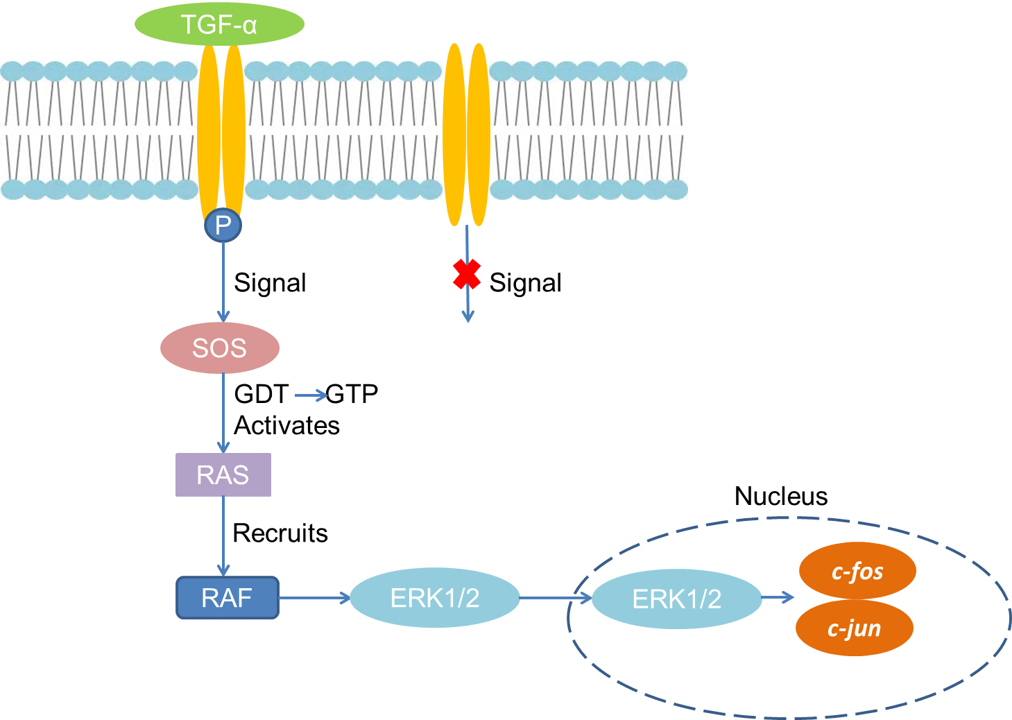 Fig 1. Mechanism of Signaling
Creative Proteomics can provide cytokine detection platform for scientific research. According to different purposes, our dedicated analysts will customize exclusive solutions for you. We aim to provide customers with high-quality and convenient services to help you accelerate the progress of your project.
Our cytokine detection service includes but is not limited to:
- One or more cytokines cytokines qualitative and quantitative detection
- Cytokines qualitative and quantitative detection of various species
- Cytokine antibodies qualitative and quantitative detection
Sample requirements
- Sample Types-Blood, serum, plasma, cell culture supernatant, cell lysate, cell culture medium, tissue homogenate, urine, tumor, etc.
- Sample Volume-It is optimal for 50 samples. This volume allows for triplicate testing of each sample.
Our advantages:
- Different detection methods can be selected based on different samples and requirements.
- Ensure the specificity and accuracy of the test by using high quality antibodies.
- Repeat the test to ensure the repeatability and accuracy of the experimental results.
- Feedback results are accurate and efficient.
Technology platform:
We mainly provide the Luminex cytokine detection platform. Luminex uses fluorescently encoded microspheres with specific antibodies to different target molecules. The different microspheres can be combined freely to a certain extent so that up to 100 analytes can be tested multiple times simultaneously in a single experiment.
The Luminex cytokine assay platform has the following advantages:
- Multiple detection: simultaneous detection of 100 biological targets
- Short experiment time: 1-3 weeks
- High sensitivity: the lower limit of accurate quantification is as low as 0.1 pg/mL
- Save samples: only need a sample volume as low as 25 μL
- Time saving: the experiment process only takes 4 hours
For your different needs, we can also provide the following detection methods:
- Enzyme-linked immunosorbent assay (ELISA)
- Flow cytometry
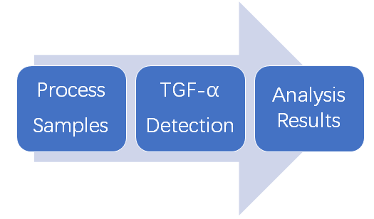
Workflow
For more information about the TGF-α detection service or need other detection requirements, please contact us.
References:
- Singh B, Coffey RJ. From wavy hair to naked proteins: The role of transforming growth factor alpha in health and disease. Semin Cell Dev Biol, 2014,28:12-21.
- Schneider MR, Sibilia M, et al. The EGFR network in bone biology and pathology. Trends EndocrinolMetab, 2009, 20(10):517-524.