
Introduction
Transforming growth factor β (TGF-β) is a group of structurally similar transforming growth factors that have multiple functions to regulate cell functions, such as regulating cell proliferation and differentiation, apoptosis, cell migration, and cell adhesion. In addition, they play important roles in embryonic development, immune monitoring, and stem cell self-renewal and differentiation. Furthermore, TGF-β plays an important role in maintaining the normal structure of the vascular wall and has a certain protective effect on atherosclerosis. In endothelial cells, TGF-β inhibits the activation of endothelial cells and reduces the recruitment of lymphocytes by inhibiting the expression of the adhesion factor E-selectin. The proliferation of vascular smooth muscle cells can be inhibited by TGF-β, which promote collagen formation, and proliferate extracellular matrix. Besides, TGF-β can affect the uptake of fat particles by macrophages, thereby reducing the formation of foam cells.
Mechanism and Function
There are five isomers of the TGF-β superfamily: TGF-β1, TGF-β2, TGF-β3, TGF-β4, and TGF-β5, which are dimeric structures consisting of 9 highly conserved cysteine residues connected by disulfide bonds. TGF-β has a wide range of biological functions in vivo and in vitro, including regulating cell growth, regulating cell phenotype, and inhibiting tumor growth. TGF-β receptors are widely expressed in tissue cells of the body, and most normal cells in the body express membrane-bound receptors of members of the TGF-β family. Since TGF-β has a highly specific affinity for its receptors, TGF-β exerts a regulatory effect on many cells. Growth inhibition is a unique property of TGF-β, it has an inhibitory effect on the proliferation of most cells, especially epithelial cells, endothelial cells, lymphoid cells and hematopoietic cells. The function expressed by TGF-β is related to the cell type. TGF-β3 plays an important role in normal wound healing in the lung, while TGF-β1 plays an important role in fibrotic wound healing in the lung. In addition, TGF-β2 mediates epithelial-mesenchymalization of the heart pad by promoting the initiation and cessation of epithelial-to-mesenchymal transition. In the nervous system, TGF-β1 has a neuroprotective effect.
The TGF-β family transmits signals mainly through Smad family proteins. According to different functions, Smad proteins are divided into 3 categories. The first category is receptor regulated Smad (R-Smad), including Smad1, Smad2, Smad3, Smad5, and Smad8; the second category is co-mediator Smad (Co-Smad), including Smad4; the third category is inhibitory Smad (I-Smad), including Smad6 and Smad7. TGF-β participates in regulating cell function by binding to TGF-β receptors on the cell surface. TGF-β binds to cell surface TGF-β receptors to phosphorylate the receptor. The phosphorylated receptor recruits and phosphorylates the downstream molecule R-Smads. In this process, the helper protein SARA is required to make R-Smad easily bind to the phosphorylated receptor. When the SSXS motif at the C-terminus of the R-Smad protein is phosphorylated, it is dissociated from the receptor complex, forms a heterodimer complex with Co-Smad. The complex is transferred into the nucleus, and regulates the target gene with transcription factors. I-Smad mainly inhibits activation of this signaling pathway by competing with R-Smad for binding to activated receptors and ubiquitinating them, degrading activated receptors, or dephosphorylation through other pathways.
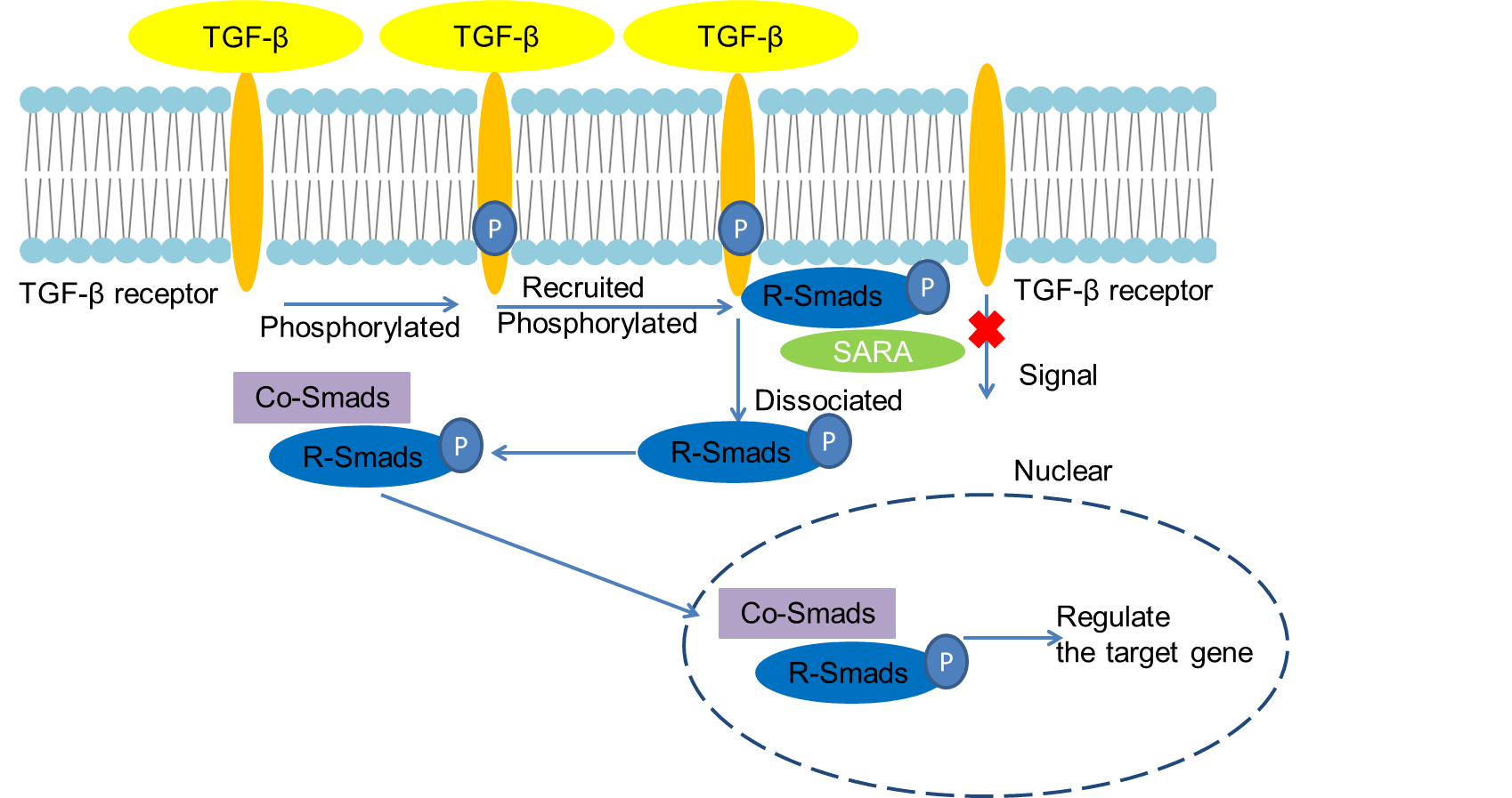 Fig 1. Mechanism of Signaling
Creative Proteomics can provide cytokine detection platform for scientific research. According to different purposes, our dedicated analysts will customize exclusive solutions for you. We aim to provide customers with high-quality and convenient services to help you accelerate the progress of your project.
Our cytokine detection service includes but is not limited to:
- One or more cytokines cytokines qualitative and quantitative detection
- Cytokines qualitative and quantitative detection of various species
- Cytokine antibodies qualitative and quantitative detection
Sample requirements
- Sample Types-Blood, serum, plasma, cell culture supernatant, cell lysate, cell culture medium, tissue homogenate, urine, tumor, etc.
- Sample Volume-It is optimal for 50 samples. This volume allows for triplicate testing of each sample.
Our advantages:
- Different detection methods can be selected based on different samples and requirements.
- Ensure the specificity and accuracy of the test by using high quality antibodies.
- Repeat the test to ensure the repeatability and accuracy of the experimental results.
- Feedback results are accurate and efficient
Technology platform:
We mainly provide the Luminex cytokine detection platform. Luminex uses fluorescently encoded microspheres with specific antibodies to different target molecules. The different microspheres can be combined freely to a certain extent so that up to 100 analytes can be tested multiple times simultaneously in a single experiment.
The Luminex cytokine assay platform has the following advantages:
- Multiple detection: simultaneous detection of 100 biological targets
- Short experiment time: 1-3 weeks
- High sensitivity: the lower limit of accurate quantification is as low as 0.1 pg/mL
- Save samples: only need a sample volume as low as 25 μL
- Time saving: the experiment process only takes 4 hours
For your different needs, we can also provide the following detection methods:
- Enzyme-linked immunosorbent assay (ELISA)
- Flow cytometry
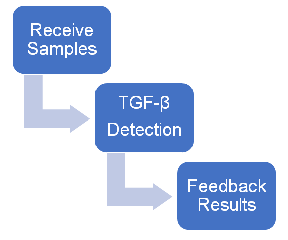
Workflow
For more information about the TGF-β detection service or need other detection requirements, please contact us.
References:
- Shi Y, Massague J. Mechanisms of TGF-beta sinaling from cell membrane to the nucleus. Cell, 2003, 113(6):685-700.
- Ungefroren H, Groth S, Sebens S, et al. Differential roles of Smad2 and Smad3 in the regulation of TGF-β1-mediated growth inhibition and cell migration in pancreatic ductal adenocarcinoma cells: control by Rac1. Mol Cancer, 2011,10(1):67.