
Introduction
Nerve growth factor (NGF) is a nerve cell growth regulator with dual biological functions of neuron nutrition and neurite-promoting growth. It can promote the growth, development, differentiation and maturation of central and peripheral neurons, and maintain nerve. The normal functioning of the system accelerates the repair of nervous system injuries. NGF is widely distributed in various tissues and organs of the body (including the brain). The concentration in the target tissue is related to the density and mRNA content of sympathetic and sensory nerve branches in the target area. NGF is the most important type in the nerve growth factor family. It exists mainly in the form of precursors in tissues, and is processed into the mature NGF in the submandibular glands. NGF contains three subunits of α, β, and γ. The active region is β subunit. The dimer composed of two 118-amino acid single chains is bonded by non-covalent bonds. It has a high degree of structure with human NGF. Homologous and biological effects have no obvious interspecific specificity. NGF is mainly distributed in the human body such as brain, ganglia, iris, heart, spleen, placenta and other tissues as well as fibroblasts, smooth muscle, skeletal muscle, glial cells, and so on.
Mechanism and Function
As the first neurotrophic factor discovered, NGF has the effect of promoting the survival and growth of nerve cells. After NGF binds to its high affinity receptor TrkA, the receptor TrkA phosphorylates to form a dimer. Phosphorylated TrkA binds to the SHC protein, and SHC acts as an adapter protein to connect to Grb containing SH2 and SH3 domains, and then binds to SOS to activate SOS. SOS activates the Ras protein, which in turn activates the RAS-induced signaling cascade. Activated Ras binds to Raf, which in turn activates MEK and ERK. After ERK enters the nucleus, it is involved in regulating cell proliferation.
NGF has a variety of biological functions. During a certain period of embryonic development, NGF is necessary for the survival of effector neurons. NGF and its receptors are widely distributed in the central nervous system. NGF produced by the hippocampus and cerebral cortex can be retrogradely transported to the basal nucleus of the forebrain through cholinergic nerves to maintain the survival and function of cholinergic neurons. In early embryonic development, the central NGF content determines the density of cholinergic nerves. In addition, when the effector neurons of NGF are damaged, such as trauma, drug damage, or even ischemia, hypoxia, neurons will undergo a series of pathological changes, including death. NGF can inhibit the effects of the above-mentioned nerve injury, mainly by inhibiting the release of toxic amino acids, inhibiting calcium ion overload, inhibiting the release of superoxide free radicals, and inhibiting apoptosis and other mechanisms to significantly reduce or prevent the occurrence of these secondary pathological damage. In addition, NGF can promote nerve regeneration and growth. Giving NGF after cutting off axons will reduce the degeneration and death of certain neurons and help increase the possibility of axon regeneration. At the same time, it also affects the time when axonal regeneration begins, the number of neurons involved in regeneration, and the quality and speed of regenerating nerves. NGF can also affect the activity of immune cells, which in turn regulates the function of the immune system. NGF inhibits the mitosis of certain tumors and promotes their benign differentiation. NGF promotes the healing response of wound tissue and promotes wound healing. NGF can also be used for the treatment of many diseases, such as delaying neurodegenerative diseases, or stimulating the growth of motor nerves in patients with spinal cord injury, accelerating burn recovery, reducing the side effects of chemotherapy and radiotherapy, treating pressure ulcers and eradicating corneal ulcers.
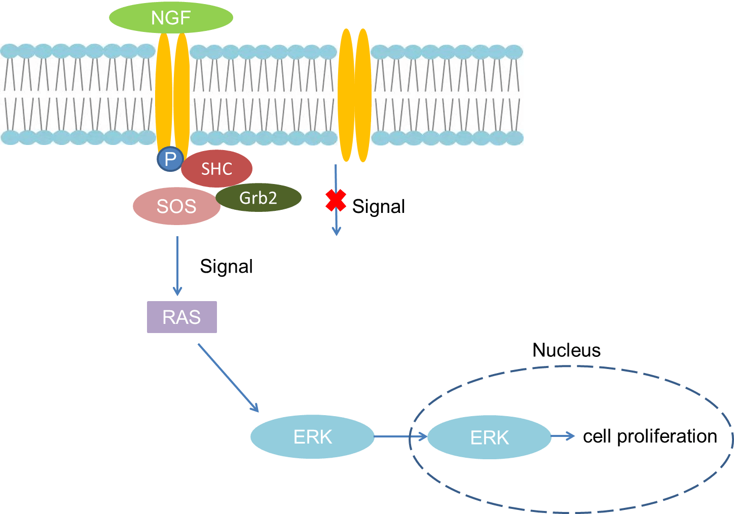 Fig 1. Mechanism of Signaling
Creative Proteomics can provide cytokine detection platform for scientific research. According to different purposes, our dedicated analysts will customize exclusive solutions for you. We aim to provide customers with high-quality and convenient services to help you accelerate the progress of your project.
Our cytokine detection service includes but is not limited to:
- One or more cytokines cytokines qualitative and quantitative detection
- Cytokines qualitative and quantitative detection of various species
- Cytokine antibodies qualitative and quantitative detection
Sample requirements
- Sample Types-Blood, serum, plasma, cell culture supernatant, cell lysate, cell culture medium, tissue homogenate, urine, tumor, etc.
- Sample Volume - It is optimal for 50 samples. This volume allows for triplicate testing of each sample.
Our advantages:
- Different detection methods can be selected based on different samples and requirements.
- Ensure the specificity and accuracy of the test by using high quality antibodies.
- Repeat the test to ensure the repeatability and accuracy of the experimental results.
- Feedback results are accurate and efficient
Technology platform:
We mainly provide the Luminex cytokine detection platform. Luminex uses fluorescently encoded microspheres with specific antibodies to different target molecules. The different microspheres can be combined freely to a certain extent so that up to 100 analytes can be tested multiple times simultaneously in a single experiment.
The Luminex cytokine assay platform has the following advantages:
- Multiple detection: simultaneous detection of 100 biological targets
- Short experiment time: 1-3 weeks
- High sensitivity: the lower limit of accurate quantification is as low as 0.1 pg/mL
- Save samples: only need a sample volume as low as 25 μL
- Time saving: the experiment process only takes 4 hours
For your different needs, we can also provide the following detection methods:
- Enzyme-linked immunosorbent assay (ELISA)
- Flow cytometry
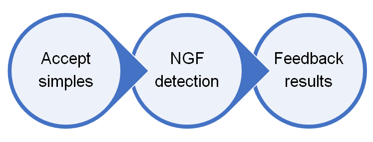
Workflow
For more information about the NGF detection service or need other detection requirements, please contact us.
References:
- Levi Montalcini R. The never growth factor: 35 years after. Science,1987,237:1154.
- Klein R, et al. The trk protooncogene encodes a receptor for nerve growth factor.Cell,1991,65:189.