
Introduction
IL-31 belongs to the IL-6 cytokine family and is mainly expressed in activated Th2 cells, especially activated helper Th2 cells, mast cells, monocytes/macrophages and dendritic cells. The main targets of IL-31 are located in the skin, lungs, intestines, and nervous system. It is also expressed at low levels in tissues such as testis, bone marrow, thymus, and spleen. IL-31 plays a major role in the induction of chronic inflammation. When the tissue is exposed to the environment, IL-31 regulates the immune process of adaptive immunity and acquired immunity. In addition, it can further stimulate the secretion of pro-inflammatory cytokines and chemokines.
Mechanism and Function
The functional receptors of IL-31 are IL-31 Receptor A (IL-31RA) and oncostatin M Receptor (OSMR), which combine to form a heterodimer complex. IL-31RA is expressed in skin tissue, brain, lung, trachea, skeletal muscle, thymus, spleen, placenta and other tissues. OSMR is more widely expressed, most in trachea, thymus and skin. In cells where OSMR and IL-31RA exist, IL-31 can activate transcription factors STAT1 and STAT3-6. But in cells with only IL-31RA or OSMR, these transcription factors did not respond to IL-31. After IL-31 binds to IL-31R, it mainly transmits cell signals through the activation of signal transducers and activators of transcription (STATs), mitogen-activated protein kinase (MAPK) and c-Jun N-terminal kinase (JNK) signaling pathways to play a biological role.
IL-31 is associated with many human diseases. It is related to the severity of atopic dermatitis, and IL-31 is found to be higher in the skin lesions of AD patients, and the patient's condition is positively correlated with the level of IL-31 in the serum. IL-31 can induce chemokine gene expression in human epidermal keratinocytes (NHEK). In NHEK, the cytokines produced by human IL-31 stimulation include CXCL1, CCL4, CCL17, CCL19, CCL22, etc., and then the expression level of IL-31 increases, leading to T cell recruitment and aggravating skin inflammation. IL-31 is also associated with allergic asthma. IL-31 can activate the JAK-STAT pathway by binding to IL-31RA on bronchial epithelial cells, thereby secreting a large amount of epidermal growth factor, vascular endothelial growth factor, monocyte chemotactic factor-1. Mast cells, eosinophils and the key cells of lung tissue autoimmunity express IL-31 and IL-31R, respectively, so that IL-31 has a direct relationship with the body's own immune system against allergens and pathogens. IL-31 and IL-31R can induce the expression of mucin 5AC (MUC5AC) gene in human respiratory epithelial cells, thereby promoting the inflammatory response of Th2 cells.
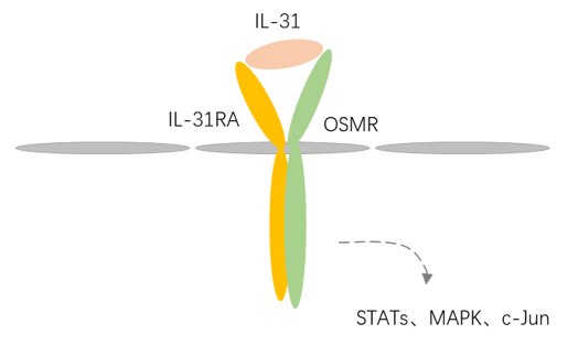 Fig 1. Mechanism of Signaling
Creative Proteomics can provide cytokine detection platform for scientific research. According to different purposes, our dedicated analysts will customize exclusive solutions for you. We aim to provide customers with high-quality and convenient services to help you accelerate the progress of your project.
Our cytokine detection service includes but is not limited to:
- Quantitative and qualitative detection of cytokines in different species
- Quantitative and qualitative detection of cytokine antibodies
- Quantitative and qualitative detection of single/mulltiple cytokines
Sample requirements
- Sample Types-Blood, serum, plasma, cerebrospinal fluid, cell culture supernatant, tissue homogenate, cell culture medium, urine, tumor, etc.
- Sample Volume-It is optimal for at least 200µl of each sample. This volume allows for triplicate testing of each sample.
Our advantages:
- Efficient design: Multiplex reaction detecting in various methods are available, which can be selected according to different samples and requirements.
- Sensitive detection: High-quality antibodies are used in the detection process to improve detection specificity and accuracy.
- Reliable results: The feedback results are professional and efficient.
Technology platform:
We mainly provide the Luminex cytokine detection platform. Luminex uses fluorescently encoded microspheres with specific antibodies to different target molecules. The different microspheres can be combined freely to a certain extent so that up to 100 analytes can be tested multiple times simultaneously in a single experiment.
The Luminex cytokine assay platform has the following advantages:
- Multiple detection: simultaneous detection of 100 biological targets
- Short experiment time: 1-3 weeks
- High sensitivity: the lower limit of accurate quantification is as low as 0.1 pg/mL
- Save samples: only need a sample volume as low as 25 μL
- Time saving: the experiment process only takes 4 hours
For your different needs, we can also provide the following detection methods:
- Enzyme-linked immunosorbent assay (ELISA)
- Flow cytometry
Workflow

For more information about the IL-31 detection service or need other detection requirements, please contact us.
References:
- Sonkoly E, Muller A, Lauerma A I, et al. IL-31: A new link between T cells and pruritus in atopic skin inflammation. Journal of Allergy & Clinical Immunology, 2006, 117(2):411-417.
- Topal F A, Zuberbier T, Makris M P, et al. The role of IL-17, IL-23 and IL-31, IL-33 in allergic skin diseases. Current Opinion in Allergy and Clinical Immunology, 2020, 20(4):367-373.