
Introduction
IL-8 is a multi-source cytokine that can be produced by monocytes, macrophages, neutrophils, fibroblasts, epithelial cells, T cells, chondrocytes, vascular endothelial cells, liver cells, etc. The main activity is to attract and activate neutrophils, further aggravate the local inflammatory response, and cause the patient's condition to worsen. Recent studies have shown that the increase in IL-8 concentration is often the pathological basis for tissue damage in certain inflammatory diseases, and it is usually the key link that leads to the development of certain diseases. The signal of IL-8 in tumor cells may increase the tumorigenicity, metastasis, invasiveness and viability of the cells. In addition, it can induce glomerular mesangial cells (GMC) to produce extracellular matrix components such as collagen fibers III, IV, and laminin.
Mechanism and Function
There are two types of IL-8 receptors, called IL-8RA and IL-8RB. IL-8A is mainly distributed in neutrophils, monocytes, T cells and melanoma cells, and has a high affinity for IL-8. , Specifically binds to IL-8. IL-8RB is a non-specific receptor. In addition to binding to IL-8R, it can also bind other CXC chemokines (GRO-α, GRO-β, GRO-γ) and neutrophil activating peptide 2, and IL- 8. GRO-α, GRO-β, GRO-γ binding has high affinity. Each chemokine binds to one or several receptors and quickly transfers to the target inflammatory cell, thereby generating an activated signal transduction pathway, which then leads to chemotaxis, proliferation, differentiation and survival of the cell.
After IL-8 binds to the receptor, it can activate Akt, protein kinase C (PKC), and cause calcium activator or mitogen-activated protein kinase (MAPK) signaling cascade. These signaling pathways can promote protein translation and regulate hypoxia-inducible factor-1 (HIF-1), nuclear factor-kappa B (NF-κB), and activator protein-1 (AAP-1), signal transducers and activators of transcription 3 (STAT3), and β-catenin activity.
IL-8 in the body is mainly a homodimer, which can activate intracellular maintenance and promote its directed migration, promote the expression of adhesion molecules by neutrophils, induce degranulation of neutrophils, and release proteolytic enzymes such as elastase, myeloperoxidase, etc. The IL-8 receptor can be expressed in cancer cells, endothelial cells, neutrophils, and tumor-associated macrophages. Therefore, IL-8 secreted by cancer cells has a great impact on the tumor environment. Inhibition of IL-8 signal in tumor cells can increase the sensitivity of tumor cells to conventional chemotherapy. IL-8 can also suppress appetite, which is related to loss of appetite in diseases. In addition, IL-8 can induce multi-gene transcription in independent factors related to tumor progression such as vascular regeneration, cell cycle management, migration and invasion, and escape.
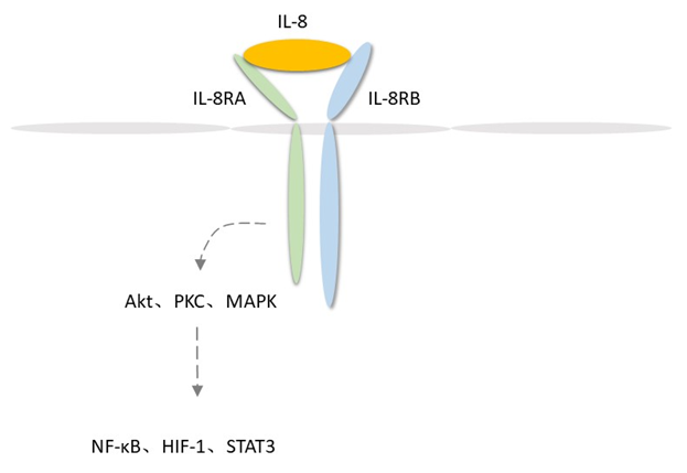 Fig 1. Mechanism of Signaling
Creative Proteomics can provide cytokine detection platform for scientific research. According to different purposes, our dedicated analysts will customize exclusive solutions for you. We aim to provide customers with high-quality and convenient services to help you accelerate the progress of your project.
Our cytokine detection service includes but is not limited to:
- Quantitative and qualitative detection of cytokines in different species
- Quantitative and qualitative detection of cytokine antibodies
- Quantitative and qualitative detection of single/mulltiple cytokines
Sample requirements
- Sample Types-Blood, serum, plasma, cerebrospinal fluid, cell culture supernatant, tissue homogenate, cell culture medium, urine, tumor, etc.
- Sample Volume - It is optimal for at least 200µl of each sample. This volume allows for triplicate testing of each sample.
Our advantages:
- Efficient design: Multiplex reaction detecting in various methods are available, which can be selected according to different samples and requirements.
- Sensitive detection: High-quality antibodies are used in the detection process to improve detection specificity and accuracy.
- Reliable results: The feedback results are professional and efficient.
Technology platform:
We mainly provide the Luminex cytokine detection platform. Luminex uses fluorescently encoded microspheres with specific antibodies to different target molecules. The different microspheres can be combined freely to a certain extent so that up to 100 analytes can be tested multiple times simultaneously in a single experiment.
The Luminex cytokine assay platform has the following advantages:
- Multiple detection: simultaneous detection of 100 biological targets
- Short experiment time: 1-3 weeks
- High sensitivity: the lower limit of accurate quantification is as low as 0.1 pg/mL
- Save samples: only need a sample volume as low as 25 μL
- Time saving: the experiment process only takes 4 hours
For your different needs, we can also provide the following detection methods:
- Enzyme-linked immunosorbent assay (ELISA)
- Flow cytometry
Workflow

For more information about the IL-8 detection service or need other detection requirements, please contact us.
References:
- Harada A , Sekido N , et al. Essential involvement of interleukin-8 (IL-8) in acute inflammation. Journal of leukocyte biology, 1994, 56(5):559.
- Kunkel S L , Standiford T , et al. Interleukin-8 (IL-8): The Major Neutrophil Chemotactic Factor in the Lung. Experimental Lung Research, 1991, 17(1):17-23.