
Introduction
Creative Proteomics can simultaneously quantitatively detect apoptosis-related cytokines and chemokines through a variety of technologies, and provide higher sensitivity and a wider detection range.
Apoptosis plays an important role in the immune system, which is closely related to intracellular homeostasis, T cell aggregation, death of reactive lymphocytes and cytotoxic response of target cells. At present, it has been found that eukaryotic cell apoptosis mainly has the following pathways: 1. Cell receptor-mediated external apoptotic pathways; 2. Internal mitochondrial pathway; 3. B-granase mediated apoptosis pathway; 4. Endoplasmic reticulum stress pathway mediates apoptosis. The detection of cytokines related to apoptosis helps to study apoptosis and the physiological or pathological processes involved.
Cytokines involved in apoptosis
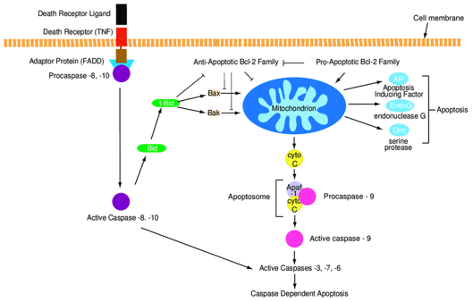
The permeabilization of the outer mitochondrial membrane (MOMP) causes the release of cytochrome C to the cytoplasm, which is mainly regulated by Bcl-2 family proteins. Under the action of intracellular apoptosis stimulating factors, Bax, Bak, etc. of Bcl-2 protein family are activated and bound to the outer membrane of mitochondria. The mitochondrial internal to cytoplasm pores are formed on the membrane, causing some proteins in the mitochondria to enter the cytoplasm through the mitochondrial membrane. The apoptotic body composed of cytochrome C, Apaf-1, and caspase-9 has the effect of cleaving the apoptotic protease caspase-7. The caspase-7 activated by lysis causes the degradation of downstream proteins related to cell life, and finally causes cell apoptosis. Other proteins that enter the cytoplasm, such as Smac/DIABLO, Omi/HtrA2, can promote the activity of caspases and thus promote cell apoptosis.
AIF is normally located in mitochondria. When cells are affected by internal apoptosis-stimulating factors, AIF can be released into the cytoplasm from mitochondria and eventually enter the nucleus, causing DNA destruction and cell death.
The transmembrane protein receptors of tumor necrosis factor (TNF) receptor superfamily members bind to related ligands and undergo oligomerization and structural changes, exposing the death domains that can bind to the junction proteins to aggregate and activate the junction proteins, triggering the activation of downstream caspases and leading to the occurrence of cell apoptosis.
FasL expressed on the surface of cytotoxic lymphocytes can bind to Fas on the surface of target cells and activate the external apoptosis pathway of target cells. In addition, cytotoxic lymphocytes can deliver some toxic particles to target cells, including TNF, B-granzyme and another perforin. TNF can mediate the external apoptosis pathway of cells. Perforin forms an inter-membrane channel on the surface of target cells, which facilitates the transfer of B-granzyme to the interior of target cells. The B-granzyme that enters the cell has a proteolytic effect, which can directly cleave and activate caspases and promote cell apoptosis.
Detectable cytokines include but are not limited to:
| AIF | APO-1/Fas | Caspase-3 | Caspase-7 | Caspase-9 |
| sCD30 | MPO | OPG | TNF-a | Cytochrome c |
Technology platform
We mainly provide the Luminex cytokine detection platform. Luminex uses fluorescently encoded microspheres with specific antibodies to different target molecules. The different microspheres can be combined freely to a certain extent so that up to 100 analytes can be tested multiple times simultaneously in a single experiment.
The Luminex cytokine assay platform has the following advantages:
- Multiple detection: simultaneous detection of 100 biological targets
- Short experiment time: 1-3 weeks
- High sensitivity: the lower limit of accurate quantification is as low as 0.1 pg/mL
- Save samples: only need a sample volume as low as 25 μL
- Time saving: the experiment process only takes 4 hours
For your different needs, we can also provide the following detection methods:
- Flow cytometry (FACS analysis): Identify and measure cell biomarkers in complex subpopulations. It can be used for intracellular cytokine detection without two hours.
- Enzyme-linked immunosorbent assay (ELISA): Use the primary antibody for capture, and conjugate the secondary antibody with an enzyme or radioisotope for detection. Our multiplexing system can detect the expression of multiple cytokines at once.
Sample preparation
- Suitable for serum, plasma, cell culture supernatant and all kinds of body fluids.
- The body fluid samples are stored in a refrigerator at -80 degrees Celsius and transported on dry ice.
Creative Proteomics can provide you with a one-stop solution. If you want to test other cytokines or other information you want to consult, please contact us. We are looking forward to cooperating with you.
Reference:
- Guerin M B, Mckernan D P, O'Brien C J, et al. Retinal ganglion cells: dying to survive. International Journal of Developmental Biology, 2002, 50(8): 665-674.