
Introduction
Interleukin 20 (IL-20) is a new member of the recently discovered interleukin 10-related cytokine family. IL-20 is a pleiotropic proinflammatory cytokine with the ability to enhance inflammation, angiogenesis and chemotaxis. Both human and mouse IL-20 molecules are composed of 176 amino acids, with a homology of 76%. IL-20 has 28% homology with IL-10, and has some homology with other IL-10 family members such as IL-19, IL-22, IL-24, IL-26 in amino acid sequence, but the biological functions are different. IL-20 can regulate the normal physiological functions of skin, activate the STAT3 pathway in keratinocytes, promote the large expression of some cytokines and chemokines, and thereby regulate the occurrence of early skin inflammation. IL-20 can also promote the proliferation and differentiation of keratinocytes, which is closely related to the pathogenesis of psoriasis.
Mechanism and Function
IL-20 is a soluble protein and belongs to the IL-10 cytokine family. IL-20 is secreted by activated keratinocytes and monocytes, and transmits signals to the cell through two receptors expressed on the surface of other endothelial cells. IL-20 has certain homology with IL-10 and other IL-10 family members in the amino acid sequence, such as 28% homology with IL-10. There are 6 conserved cysteine residues in the IL-20 molecule, of which cys81 and cys134 form a disulfide bond, but do not form a dimer. IL-20 can play a role by binding to the receptor, and two subunits must exist at the same time to efficiently bind to the ligand. IL-20 receptors (IL-20Rα/IL-22Rα, IL-20Rβ) are expressed in renal tissue interstitial fibroblasts, renal epithelial cells, mesangial cells and podocytes. After IL-20 binds to the receptor, it activates STAT3 by binding to JAK1 and TYK2. The two STAT3 form a homodimer through the nuclear membrane to enter the nucleus, regulate gene expression, and produce biological effects.
IL-20 can promote the secretion of cytokines and chemokines. Under the stimulation of IL-20, the expressions of TNF-α, MCP-1 and MRP-14 in cells were up-regulated, and these cytokines are closely related to the early inflammation initiation and immune response. Moreover, IL-20 can also stimulate the proliferation and differentiation of keratinocytes, which may be related to T cells. In addition, IL-20 can imbalance the pro-apoptosis and pro-survival molecules, induce programmed death of renal epithelial cells, and finally activate the bypass of mitochondrial apoptosis. IL-20 is an important medium for regulating renal tubular cell apoptosis and promoting renal fibrosis, and stimulates mesangial cells to produce iNOS, ROS, MCP-1, and IL-6. And iNOS promotes the production of more nitric oxide, causing kidney tissue damage. In addition, IL-20 also induced the production of TGF-β1, matrix metalloproteinase-2 (MMP-2) and MMP-9 mesangial cells. These results indicate that IL-20 may be involved in renal fibrosis. Infiltrating inflammatory cells, mesangial cells, or renal epithelial cells produce IL-20, which then triggers fibroblasts to produce more fibrotic factors. IL-20 also induces epithelial cell apoptosis in an autocrine or paracrine manner. Blocking the function of IL-20, including monoclonal antibodies or small molecules, may be a viable treatment option for IL-20-mediated kidney injury, which can delay its progress and reduce its severity.
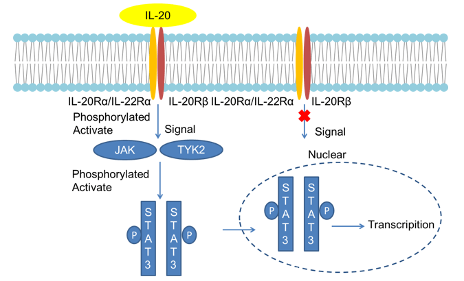 Fig 1. Mechanism of Signaling
Creative Proteomics can provide cytokine detection platform for scientific research. According to different purposes, our dedicated analysts will customize exclusive solutions for you. We aim to provide customers with high-quality and convenient services to help you accelerate the progress of your project.
Our cytokine detection service includes but is not limited to:
- One or more cytokines qualitative and quantitative detection
- Cytokines qualitative and quantitative detection of various species
- Cytokine antibodies qualitative and quantitative detection
Sample requirements
- Sample Types-Blood, serum, plasma, cell culture medium, tissue homogenate, etc.
- Sample Volume - It is optimal for 50 samples. This volume allows for triplicate testing of each sample.
Our advantages:
- Different detection methods can be selected based on different samples and requirements.
- Ensure the specificity and accuracy of the test by using high quality antibodies.
- Repeat the test to ensure the repeatability and accuracy of the experimental results.
- Feedback results are accurate and efficient.
Technology platform:
We mainly provide the Luminex cytokine detection platform. Luminex uses fluorescently encoded microspheres with specific antibodies to different target molecules. The different microspheres can be combined freely to a certain extent so that up to 100 analytes can be tested multiple times simultaneously in a single experiment.
The Luminex cytokine assay platform has the following advantages:
- Multiple detection: simultaneous detection of 100 biological targets
- Short experiment time: 1-3 weeks
- High sensitivity: the lower limit of accurate quantification is as low as 0.1 pg/mL
- Save samples: only need a sample volume as low as 25 μL
- Time saving: the experiment process only takes 4 hours
For your different needs, we can also provide the following detection methods:
- Enzyme-linked immunosorbent assay (ELISA)
- Flow cytometry
Workflow

For more information about the IL-20 detection service or need other detection requirements, please contact us.
References:
- Blumberg H, et al. Interleutin 20: Discovery, receptor identification, and role in epidermal function. Cell, 2001, 104(1): 9-19.
- Dumoutier L, et al. STAT activation by IL-19, IL-20 and mda-7 through IL-20 receptor complexes of two types. J Immnol, 2001, 167(7): 3545-3549.