
Introduction
Insulin-like growth factor (IGF) is a group of growth-promoting peptides. Its secreting cells are widely distributed in human liver, kidney, lung, heart, brain, and intestine. The production of IGF-I is more dependent on GH, which has a strong growth-promoting effect and is an important growth factor in childhood. IGF-I synthesized in various tissues mostly exerts its growth-promoting effects in an autocrine or paracrine manner, while IGF-I synthesized by the liver enters the blood circulation and acts on target cells in an endocrine manner. The level of IGF-I in the body is regulated by GH. IGF- I also has a negative feedback regulation effect on GH secretion. IGF-II is more insulin-like and plays an important role in fetal growth. IGF receptors are divided into two types, namely, IGF-I receptor (IGF-I R) and IGF-II receptor (IGF-II R). The former is a tyrosine kinase-type receptor and is composed of two α subunits. The latter has no amino acid kinase activity.
Mechanism and Function
IGF is a type of multifunctional cell regulatory factor. Most IGFs are synthesized in the liver and mainly exist in the blood circulation. IGF mediates growth hormone stimulation, regulates tissue growth and development, and plays an important role in muscle volume and strength, maintenance of body composition, and regulation of nutrient metabolism. Moreover, IGF may be an important autocrine and paracrine signaling molecule during the development of the central nervous system. IGF-I is considered to be a survival factor for glial progenitor cells and oligodendrocytes. Both IGF-I and IGF- II can be used as muscle-derived neurotrophic factors to stimulate the growth of intramuscular neurites. IGF exerts its biological effects through the endocrine, paracrine, and / or autocrine methods, and is mediated by the target cell surface receptors IGF-I R and IGF-II R, and is regulated by IGF-binding proteins. Most extrahepatic tissues can produce IGF locally, and most of them are not regulated by GH and play an important role in local tissues.
At present, there are mainly two cellular signal transduction pathways, namely the RAS / RAF / MEK / ERK signal transduction pathway and the PI3K / AKT signal transduction pathway. Studies have shown that IGF-mediated mitosis and cell proliferation and differentiation are mainly accomplished through the RAS / RAF / MEK / ERK signal transduction pathway. When IGFR binds to IGF, its tyrosine and serine residues are phosphorylated, which in turn activates IGFR. The activated IGFR binds to the SH2 domain of growth factor receptor binding protein-2 (Grb2), while the SH3 domain of Grb2 can bind to the son of sevenless (SOS), which results RAS dissociates GDP and binds GTP, thereby activating RAS. Activated RAS further binds to the amino terminus of the serine / threonine protein kinase RAF-1, thereby activating RAF-1. RAF-1 activates MEK by phosphorylating MEK1 / MEK2. MEK is a bispecific kinase that can phosphorylate serine / threonine and tyrosine, and ultimately selectively activate ERK1 and ERK2. ERK is a proline-directed serine / threonine kinase that can phosphorylate serine / threonine adjacent to proline. After mitogen stimulation, ERK receives upstream cascade response signals, which can be converted into the nucleus and regulate cell proliferation and differentiation.
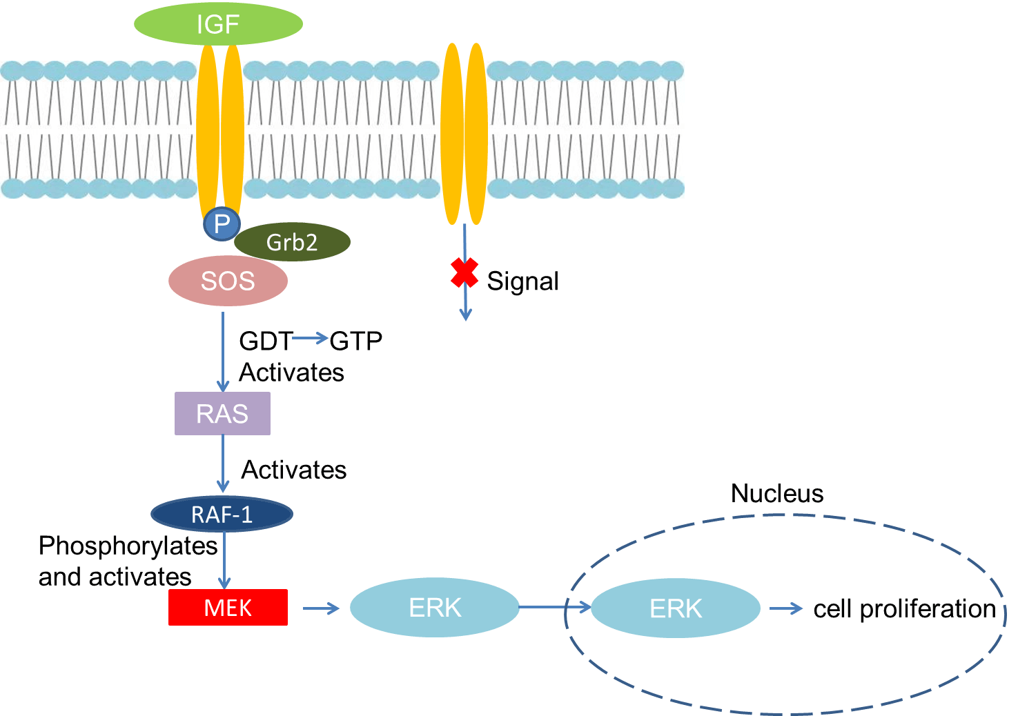 Fig 1. Mechanism of Signaling
Creative Proteomics can provide cytokine detection platform for scientific research. According to different purposes, our dedicated analysts will customize exclusive solutions for you. We aim to provide customers with high-quality and convenient services to help you accelerate the progress of your project.
Our cytokine detection service includes but is not limited to:
- One or more cytokines cytokines qualitative and quantitative detection
- Cytokines qualitative and quantitative detection of various species
- Cytokine antibodies qualitative and quantitative detection
Sample requirements
- Sample Types-Serum, cell supernatant, urine, body fluids, lavage fluid, cerebral spinal cord, atrial fluid, pleural fluid, tissue, etc.
- Sample Volume - It is optimal for at least 200µl of each sample. This volume allows for triplicate testing of each sample.
Our advantages:
- Different detection methods can be selected based on different samples and requirements.
- Ensure the specificity and accuracy of the test by using high quality antibodies.
- Repeat the test to ensure the repeatability and accuracy of the experimental results.
- Feedback results are accurate and efficient
Technology platform:
We mainly provide the Luminex cytokine detection platform. Luminex uses fluorescently encoded microspheres with specific antibodies to different target molecules. The different microspheres can be combined freely to a certain extent so that up to 100 analytes can be tested multiple times simultaneously in a single experiment.
The Luminex cytokine assay platform has the following advantages:
- Multiple detection: simultaneous detection of 100 biological targets
- Short experiment time: 1-3 weeks
- High sensitivity: the lower limit of accurate quantification is as low as 0.1 pg/mL
- Save samples: only need a sample volume as low as 25 μL
- Time saving: the experiment process only takes 4 hours
For your different needs, we can also provide the following detection methods:
- Enzyme-linked immunosorbent assay (ELISA)
- Flow cytometry
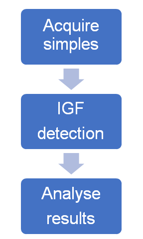
Workflow
For more information about the IGF detection service or need other detection requirements, please contact us.
References:
- Dalle S, Imamura T, et al.Insulin induces heterologous desensitization of G-protein-coupled receptor and insulin-like growth factor I signaling by downregulating beta-arrestin-1. MOL Cell Biol, 2002, 22(17) : 6272-6285.
- Deborah H. Anderson. Role of lipids in the MAPK signaling pathway. Prog Lipid Res. 2006, 18: 1-18.