Introduction
IL-5 was first discovered in T cell conditioned medium in 1980. It can replace T cells to cooperate with thymus-dependent antigen antibody response in vitro, which is called T cell replacing factor (TRF). In addition, IL-5 has important regulatory effects on the proliferation and differentiation of B cells and eosinophils. It is also called B cell growth factor-II (BCGF-Ⅱ), IgA-enhancing factor (IgA-EF), eosinophilcolony-stimulating factor (Eo-CSF) and eosinophil differentiation factor (EDF). Human and mouse IL-5 have 70% homology at the amino acid level, and biological effects have cross-reactivity. In humans, IL-5 is mainly produced by activated T cells. In mice, it is produced by Th2 cells.
Mechanism and Function
Mouse IL-5 is composed of 133 amino acid residues and contains a 21 amino acid signal peptide. The mature IL-5 molecule contains 112 amino acid residues, and the molecular chain is 12-15 kDa. There are 3 glycosylation sites, and the molecular weight after carbonylation is 18kDa. Glycosylation plays an important role in IL-5 activity expression and binding of corresponding receptors. Mouse IL-5 usually exists as a disulfide-linked dimer with a molecular weight of 45 kDa. Human IL-5 is composed of 134 amino acid residues, contains a signal state of 22 amino acids, and has 2 glycosylation sites. Mouse IL-5 gene is located on chromosomes 5 and 11 respectively, and is closely linked to the genes of IL-3, IL-4, and GM-CSF hematopoietic factors.
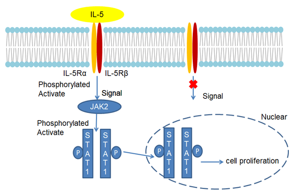
The mouse IL-5 receptor is composed of two chains of α and β. The α chain is a glycoprotein containing 415 amino acids, 17 amino acids in the leader sequence, 322 amino acid residues in the outer membrane region, 22 amino acid residues in the empty membrane regions, and 54 amino acid residues in the cytoplasm. The alpha chain alone has low affinity for IL-5 and can participate in signal transduction. The beta chain does not bind IL-5 alone, and together with the alpha chain constitutes a high affinity receptor. Mouse IL-5Rβ is the same as the IL-3R β chain, and human IL-5Rβ is common to the IL-3Rβ chain and GM-CSFRβ. Due to different mRNA splicing, two soluble mouse IL-5Rα chains have been found, one of which inhibits IL-5 binding to IL-5R. Furthermore, human and mouse IL-5Rα chains have 79% homology. In eosinophils, IL-5 activates Jak2-Stat. Stat exists in the cytoplasm in an inactive form. After being activated by Jak, it forms a dimer and transfers to the nucleus to bind to 5'-TTCNNNGAA-3 ', a conserved region of GAS elements, and plays a role in regulating transcription of cytokine-specific genes.
The biological functions of IL-5 mainly include 4 aspects. First, IL-5 promotes antigen-stimulated B cells to differentiate into antibody-synthesizing cells, which mainly acts on B cells entering the late stage of cell appreciation and increases the expression of IL-2R in B cells. This stimulation has similar functions to IL-6. IL-5 only works for a short time after B cell stimulation. Second, IL-5 promotes IgA synthesis. As an IgA-specific promoter, mIgM-positive B cells are differentiated into mIgA-positive B cells. In addition, IL-5 acts on IgA-type B cells, promotes their proliferation and differentiation, and becomes a plasma cell that secretes IgA. IL-4 has the synergistic effect of IL-5 to promote IgA synthesis, and IL-5 also promotes the secretion of IgM. Third, IL-5 cooperates with ConA or IL-2 to induce the differentiation of killer T cell precursors (CTPp) into CTL in the thymus. Fourth, IL-5 chemotaxis human eosinophils, prolong the survival time of mature eosinophils, stimulate the function of human and mouse eosinophils, and induce the differentiation of eosinophils.
 Fig 1. Mechanism of Signaling
Creative Proteomics can provide cytokine detection platform for scientific research. According to different purposes, our dedicated analysts will customize exclusive solutions for you. We aim to provide customers with high-quality and convenient services to help you accelerate the progress of your project.
Our cytokine detection service includes but is not limited to:
- One or more cytokines qualitative and quantitative detection
- Cytokines qualitative and quantitative detection of various species
- Cytokine antibodies qualitative and quantitative detection
Sample requirements
- Sample Types-Blood, serum, plasma, cell culture medium, tissue homogenate, etc.
- Sample Volume - It is optimal for 50 samples. This volume allows for triplicate testing of each sample.
Our advantages:
- Different detection methods can be selected based on different samples and requirements.
- Ensure the specificity and accuracy of the test by using high quality antibodies.
- Repeat the test to ensure the repeatability and accuracy of the experimental results.
- Feedback results are accurate and efficient.
Technology platform:
We mainly provide the Luminex cytokine detection platform. Luminex uses fluorescently encoded microspheres with specific antibodies to different target molecules. The different microspheres can be combined freely to a certain extent so that up to 100 analytes can be tested multiple times simultaneously in a single experiment.
The Luminex cytokine assay platform has the following advantages:
- Multiple detection: simultaneous detection of 100 biological targets
- Short experiment time: 1-3 weeks
- High sensitivity: the lower limit of accurate quantification is as low as 0.1 pg/mL
- Save samples: only need a sample volume as low as 25 μL
- Time saving: the experiment process only takes 4 hours
For your different needs, we can also provide the following detection methods:
- Enzyme-linked immunosorbent assay (ELISA)
- Flow cytometry
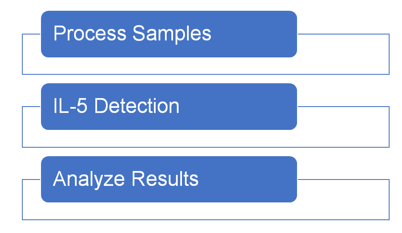
Workflow

For more information about the IL-5 detection service or need other detection requirements, please contact us.
References:
- Fabrice G, et al. EMBO J,1995, 14:2005-2013.
- Tjomme V B, et al. Blood, 1995,85:1442-1448.

