
- Service Details
- Case Study
Infectious Inflammation and Cytokine
Redness, hot pain as well as sepsis, are symptoms of the pathological process of infectious inflammation, a phenomenon that occurs with the participation of mediators produced by a large number of inflammatory cells, where cytokines are also involved. Cytokines are biologically active low molecular weight proteins or peptides secreted by various cells that regulate cell growth and differentiation, immune function, inflammation and wound healing. When pathogens invade or the body ages, inflammatory cytokines activate the body's immune system to defend against invaders. And when invaders are eliminated and the body needs to regain stability, anti-inflammatory cytokines are secreted. The constant interaction and balance between inflammatory and anti-inflammatory cytokines is an important part of the human immune system.
Pro-Inflammatory and Anti-Inflammatory Pathways
Pro-inflammatory cytokines are endogenous peptides with powerful biological effects produced mainly by cells of the immune system, including interleukin-1β, tumor necrosis factor (TNF-α), IL-6, IL-18, IL-17 and IL-15, etc. Among them, IL-1 is capable of activating a variety of pro-inflammatory cytokines of immune and inflammatory cells, and it includes IL-1α and IL-1β. IL-1β promotes the proliferation and differentiation of B cells, mediates the secretion of immunoglobulins, and enhances the process of cellular and humoral immune-mediated tissue injury. Another typical pro-inflammatory cytokine TNF-α can stimulate macrophages, fibroblasts, smooth muscle cells, epithelial cells and endothelial cells to cause arachidonic acid metabolites and proteases, etc. It can also phagocytose cell products and complement fragments to cause cell necrosis and edema, leading to tissue damage of untouched cells. Besides, it can promote the secretion of intestinal epithelial cells and the expression of IL-8 gene, upregulate T Number of cells, eosinophils and basophils.
Inflammatory cytokines can regulate the response of pro-inflammatory cytokine production, mainly including IL-4, IL-5, IL-10, IL-13, transforming growth factor (TGF), epiodermal growth factor and so on. Among them, IL-4 can inhibit the production of other pro-inflammatory cytokines such as IL-1 and IL-6, as well as the production of lymphocytes and macrophages. IL-10 can inhibit the production of activated monocytes, macrophages, granulocytes and T cells. And IL-13 can inhibit the production of pro-inflammatory transmitters and down-regulate the function of monocytes with cytotoxicity. TGF-β can inhibit the inflammatory response of intestine. And EGF can stimulate tissue repair and protect the mucosa of gastrointestinal tract.
Creative Proteomics can provide cytokine detection platform for scientific research. According to different purposes, our dedicated analysts will customize exclusive solutions for you. We aim to provide customers with high-quality and convenient services to help you accelerate the progress of your project.
Comprehensive Inflammation Cytokine Detection Services at Creative Proteomics
Cytokine Profiling: We provide cytokine profiling services using the advanced Luminex multiplex assay, allowing for the simultaneous detection and quantification of multiple cytokines in your samples.
Cytokine Quantification: Creative Proteomics can accurately quantify individual cytokines of interest using various quantitative methods.
Cytokine Biomarker Discovery: Our experts can assist you in identifying novel cytokine biomarkers associated with inflammation, providing valuable insights into disease mechanisms.
Cytokine Signaling Pathway Analysis: We offer services to investigate the signaling pathways involved in cytokine-mediated inflammation, helping you understand the underlying molecular mechanisms.
Customized Assays: If you have specific requirements or unique cytokines to detect, we can develop customized assays tailored to your research needs.
Quality Assurance: Our services are performed with the highest standards of quality assurance to ensure the accuracy and reliability of your data.
Technology Platform for Inflammation Cytokine Assay
We mainly provide the Luminex cytokine detection platform. Luminex uses fluorescently encoded microspheres with specific antibodies to different target molecules. The different microspheres can be combined freely to a certain extent so that up to 100 analytes can be tested multiple times simultaneously in a single experiment.
The Luminex cytokine assay platform has the following advantages:
- 96-well plate design for batch detection of multiplexed factors up to 80 samples
- High sensitivity, as low as pg/mL
- Multi-factor assays performed with 50 μL liquid sample and 200 μg total protein
- Maximized information in a single sample and cross-analysis allowed between analytes
For your different needs, we can also provide the following detection methods:
- Enzyme-linked immunosorbent assay (ELISA)
- Flow cytometry
List of Customizable Inflammation Cytokines Analysis
| GM-CSF | IFN-γ | IL-1β | IL-2 | IL-4 |
| IL-5 | IL-6 | IL-8 | IL-10 | IL-12 |
| IL-13 | IL-15 | IL-17 | IL-18 | IL-23 |
| IL-27 | IL-33 | IP-10 | MCP-1 | MIP-1α |
| MIP-1β | RANTES | TGF-β | TNF-α | Eotaxin |
Sample Requirements for Inflammation Cytokine Assay
| Sample Type | Storage Conditions | Additional Notes |
|---|---|---|
| Serum | Store at -80°C or -20°C | Avoid repeated freeze-thaw cycles. |
| Plasma | Store at -80°C or -20°C | Avoid hemolysis during sample collection. |
| Cerebrospinal Fluid | Store at recommended temperature | Specify any special handling instructions if applicable. |
| Tissues | Snap-freeze in liquid nitrogen | Include information about tissue type and preservation method. |
| Cell Culture Supernatant | Store at -80°C or -20°C | Provide detailed information about cell culture conditions. |
| Urine | Store at recommended temperature | Provide information about sample collection and preservation. |
| Other Biological Fluids | As appropriate | Please contact us for specific requirements. |
Applications of Inflammation Cytokine Analysis
Exploring Immune Responses:
Inflammation cytokines play a pivotal role in the body's immune response. Our services enable you to delve into the intricate web of cytokine interactions, helping you understand the underlying mechanisms involved in various inflammatory processes. Whether you're studying cellular signaling pathways, immune system modulation, or host-pathogen interactions, our analysis can provide valuable insights.
Biomarker Discovery:
Identifying reliable biomarkers is crucial for many research endeavors. Our Inflammation Cytokine Analysis Services allow you to screen a wide array of cytokines simultaneously, facilitating the discovery of potential biomarkers associated with specific inflammatory conditions or diseases. These biomarkers can aid in disease monitoring and risk assessment.
Drug Development Support:
For pharmaceutical researchers, understanding cytokine profiles is essential in the development of targeted therapies and drug candidates. Our services assist in characterizing cytokine responses to potential drug compounds, helping you make informed decisions during drug development stages.
Immunology Research:
In the field of immunology, the ability to measure multiple cytokines in a single sample can be a game-changer. Our technology enables you to profile immune responses comprehensively, allowing you to investigate the effects of different stimuli on cytokine production and immune cell activation.
Preclinical Models and Epidemiological Studies:
Researchers working with preclinical models or conducting epidemiological studies can benefit from our Inflammation Cytokine Analysis Services. These studies often involve the assessment of cytokine profiles to gain insights into disease progression, treatment efficacy, or the impact of environmental factors on immune responses.
For more information about cytokines in inflammation arthritis detection service or need other detection requirements, please contact us.
References:
- Zheng K Y . The Biological Functions of T Helper 17 Cell Effector Cytokines in Inflammation. Immunity, 2008.
- Ouyang W, Rutz S, Crellin N K, et al. Regulation and Functions of IL-10 Family Cytokines in Inflammation and Diseases. Annual Review of Immunology, 2010, 29(1):71-109.
Case Cytokine Signature Associated with Disease Severity in Dengue
Background
Dengue is a rapidly spreading viral disease transmitted by Aedes mosquitoes, with a significant global health impact. The study aimed to investigate the cytokine signature associated with disease severity in dengue, considering the role of dengue virus serotypes and host immune responses.
Samples
The study included 98 patients who presented during a dengue outbreak in eastern India in 2016. These patients were categorized into groups based on disease severity, including dengue fever (with and without warning signs) and severe dengue cases. Age and sex-matched healthy control subjects were also included for comparison.
Technical Methods
Detection of Dengue Virus
The existence of the dengue virus (DENV) was determined using multiple techniques, encompassing NS1 antigen detection, IgM capture ELISA, and serotype-specific RT-PCR. This comprehensive approach facilitated the identification of the specific DENV serotypes circulating within the studied population.
Cytokine Analysis
Cytokine analysis primarily relied on the utilization of the multiplex Luminex assay, a powerful method allowing for the concurrent quantification of numerous cytokines and chemokines from a single plasma sample. The workflow of this assay involved the following key steps:
a. Bead-Based Technology: The assay harnessed magnetic beads, each coated with antibodies specific to distinct cytokines. These beads were uniquely labeled with fluorescent dyes of different colors, enabling their differentiation.
b. Plasma Incubation: A small volume of plasma extracted from each study subject was introduced to a set of beads, with each bead tailored to a different cytokine. As plasma inherently contains cytokines, they selectively adhered to their corresponding antibody-coated beads.
c. Washing: Subsequent to incubation, thorough washing of the samples was performed to eliminate unbound proteins and potential contaminants.
d. Detection: The beads, now laden with captured cytokines, were processed through a flow cytometer. This specialized instrument recognized the individual beads based on their unique fluorescence and gauged the intensity of the emitted fluorescence, directly correlated to the quantity of captured cytokines.
e. Data Analysis: A dedicated computer program meticulously analyzed the data generated by the flow cytometer. It computed the concentrations of multiple cytokines present in the plasma sample, yielding a comprehensive and informative cytokine profile for each study subject.
Statistical Analysis
Statistical analysis was an integral part of the study, aiming to draw meaningful comparisons between cytokine levels in dengue patients and the healthy control group. Various statistical tests were employed to gauge the significance of observed differences. These tests encompassed Student's t-test, ANOVA, and Fisher's exact test. Additionally, proportions were computed, accompanied by the determination of 95% confidence intervals to provide a robust understanding of the data's variability. To delve deeper into the data and uncover intricate relationships, multivariate analyses were conducted. These analyses included Principal Component Analysis (PCA) and Partial Least Squares Discriminant Analysis (PLS-DA). The application of these sophisticated techniques aimed to discern discernible cytokine patterns that might be linked to disease severity and specific dengue serotypes.
In essence, the utilization of a diverse array of statistical approaches allowed for a comprehensive exploration of the cytokine data, enhancing the study's ability to identify meaningful associations and patterns within the dataset.
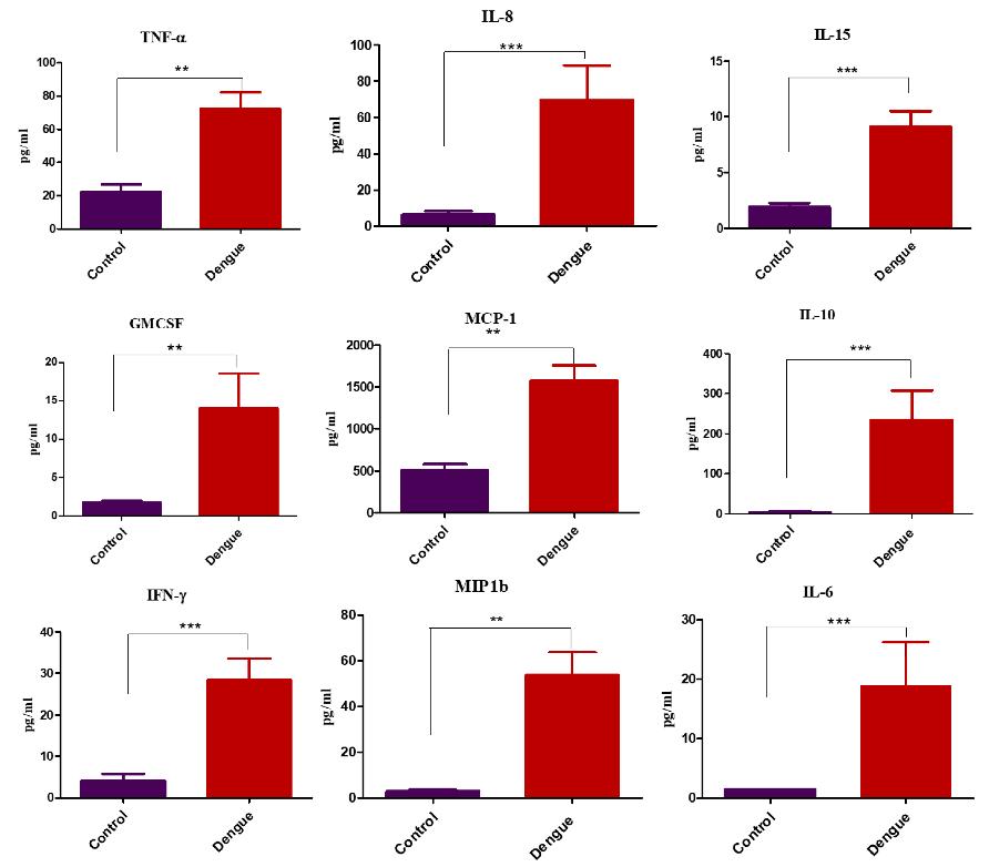 Cytokine levels in healthy controls and dengue positive subjects.
Results
During the outbreak, all four serotypes of Dengue Virus (DENV-1, DENV-2, DENV-3, and DENV-4) were detected, with DENV-2 being the prevailing serotype in a majority, accounting for 55% of cases.
Comparing dengue patients to healthy controls, elevated levels of inflammatory cytokines and chemokines were discerned. These included GM-CSF, IFN-γ, IL-10, IL-15, IL-8, MCP-1, IL-6, MIP-1β, and TNF-α, signifying a heightened immune response in dengue patients.
Significantly elevated levels of four specific cytokines, namely IFN-γ, GM-CSF, IL-10, and MIP-1β, were observed in patients with severe dengue in contrast to those with mild dengue fever. This observation suggests the potential utility of these cytokines as predictive markers for assessing disease severity.
To distinguish between DENV-2 and other DENV serotypes based on cytokine profiles, advanced multivariate analyses such as Principal Component Analysis (PCA) and Partial Least Squares Discriminant Analysis (PLS-DA) were employed. These analyses shed light on serotype-specific immune responses, revealing valuable insights into the immune reactions triggered by different DENV serotypes.
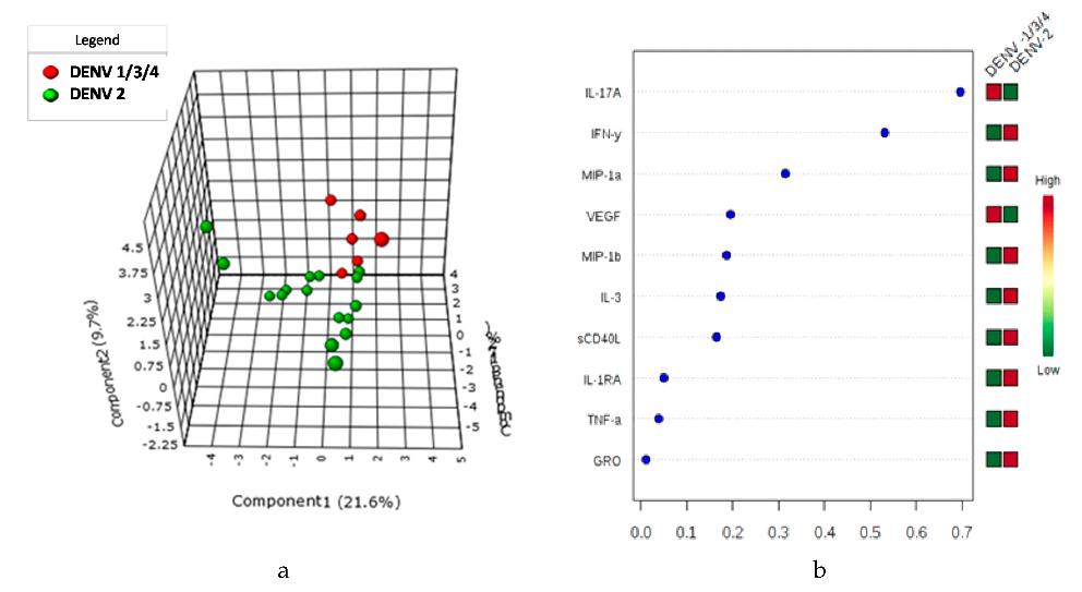 sPLS-DA and VIP score of Cytokine expression profile in DENV-2 vs DENV -1, -3, -4 serotypes in Dengue Patients.
Reference:
- Patro, A. Raj Kumar, et al. "Cytokine signature associated with disease severity in dengue." Viruses 11.1 (2019): 34.