
Introduction
IL-4 is a cytokine secreted by type 2 T helper cells, mast cells, and basophils. It has an immune function and is an anti-inflammatory factor. Besides, it also plays a central role in regulating antigen-stimulated initial T cell differentiation, causing T cells to produce cytokines such as IL-10, IL-14, and inhibiting CD4+ T cells from secreting IFN-γ. It can only exert its biological activity if it binds to a specific receptor on the membrane of the target cell.
Mechanism and Function
IL-4 is a dense, hydrophobic, spherical protein consisting of 129 amino acid residues. The biological effects of IL-4 are mediated by binding to its receptors, which are distributed in T cells, B cells, thymocytes, macrophages, bone marrow cells, macrophages and other cells. After binding to its receptor IR-4R, it can play a variety of biological functions, such as participating in the differentiation of primitive helper T cells, clearing parasites in the body, and triggering hypersensitivity. There are two types of IL-4 receptor (IL-4R): type I receptor complex IL-4Rα / IL-2Rγc and type II receptor complex IL-4Rα / IL-13Rα1. Type I receptors are mainly expressed on the surface of hematopoietic cells, and type II is often expressed on the surface of other cells such as non-hematopoietic cells and tumor cells. IL-4 can bind to IL-4R on the surface of tumor cells and activate the JAK / STAT6 signaling pathway. When IL-4 binds to IL-4Rα on the cell membrane, JAK enzyme is activated, and then STAT6 in the cytoplasm is phosphorylated and activated to form a STAT6 homodimer, thereby entering the nucleus to bind to specific DNA sites and induce transcriptional expression of IL-4 and other related inflammatory genes to amplify the inflammatory response and cause fibrotic degeneration of diseased tissue. Among T lymphocytes, only IL-4 can induce the activation of STAT6, and phosphorylated activation of STAT6 is necessary for many functions of IL-4, such as causing the body's immune response, or directly affecting tumor cells, causing it to escape. Due to this mechanism of the IL-4/IL-4R signaling pathway, and type II IL-4R is overexpressed in many solid tumors, such as gastric cancer, lung cancer, and kidney cancer. So IL-4R has a lot to do with tumors.
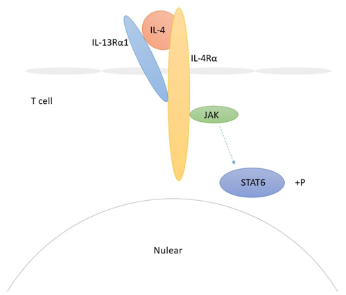 Fig 1. Mechanism of Signaling
Creative Proteomics can provide cytokine detection platform for scientific research. According to different purposes, our dedicated analysts will customize exclusive solutions for you. We aim to provide customers with high-quality and convenient services to help you accelerate the progress of your project.
Our cytokine detection service includes but is not limited to:
- Quantitative and qualitative detection of cytokines in different species
- Quantitative and qualitative detection of cytokine antibodies
- Quantitative and qualitative detection of single/multiple cytokines
Sample requirements
- Sample Types-Blood, serum, plasma, cerebrospinal fluid, cell culture supernatant, tissue homogenate, cell culture medium, urine, tumor, etc.
- Sample Volume - It is optimal for at least 200µl of each sample. This volume allows for triplicate testing of each sample.
Our advantages:
- Efficient design: Multiplex reaction detecting in various methods are available, which can be selected according to different samples and requirements.
- Sensitive detection: High-quality antibodies are used in the detection process to improve detection specificity and accuracy.
- Reliable results: The feedback results are professional and efficient.
Technology platform:
We mainly provide the Luminex cytokine detection platform. Luminex uses fluorescently encoded microspheres with specific antibodies to different target molecules. The different microspheres can be combined freely to a certain extent so that up to 100 analytes can be tested multiple times simultaneously in a single experiment.
The Luminex cytokine assay platform has the following advantages:
- Multiple detection: simultaneous detection of 100 biological targets
- Short experiment time: 1-3 weeks
- High sensitivity: the lower limit of accurate quantification is as low as 0.1 pg/mL
- Save samples: only need a sample volume as low as 25 μL
- Time saving: the experiment process only takes 4 hours
For your different needs, we can also provide the following detection methods:
- Enzyme-linked immunosorbent assay (ELISA)
- Flow cytometry
Workflow

For more information about the IL-4 detection service or need other detection requirements, please contact us.
References:
- Jiang H, et al. IL-4/IL-13 signaling beyond JAK/STAT. Journal of Allergy and Clinical Immunology, 2000, 105(6):1063-1070.
- Takeda K, et al. Essential role of Stat6 in IL-4 signalling. Nature (London), 1996, 380(6575):627-630.