
Introduction
Fibroblast growth factor (FGF) is a peptide secreted by the pituitary and hypothalamus, including acidic (pI 5.6) and basic (pI 9.6). Acidic FGF (aFGF) and basic FGF (bFGF) are the prototype FGF members named for their different isoelectric points. Acidic FGF is highly expressed in the brain, retina, bone matrix, and osteosarcoma. Basic FGF is found in a variety of tissues, including the pituitary gland, nerve tissue, adrenal cortex, corpus iuteum, and placenta. Acidic and basic FGFs stimulate the proliferation of mesoderm-derived cells, neuroectodermal cells, ectodermal cells, and endoderm-derived cells. They have chemotactic and mitotic effects on endothelial cells and induce the release of substances that damage the basement membrane. These two FGFs and other members of the FGF family play important roles in regulating cell proliferation, migration, differentiation, and angiogenesis.
Mechanism and Function
FGFs are effective regulators of cell proliferation, differentiation, and function, and play important roles in normal development, tissue maintenance, wound repair, and angiogenesis. FGFs are also associated with several pathological conditions. FGF gene mutations have been linked to various diseases including cancer, cardiovascular disease, osteoarthritis, diabetes, Parkinson's disease, and hypophosphatemia. The human FGF family includes 22 members, FGF-1 to FGF-23 (except FGF-15). With the exception of four members (FGF11, FGF12, FGF13, and FGF14), all FGFs bind to the FGF receptor (FGFR) and are divided into six different FGF subfamilies3. FGF family members generally have 30-50% amino acid sequence homology, two conserved cysteine residues, and strong binding affinity to heparin. FGF activates FGFRs by binding to FGFRs. Activated FGFRs transduce signals through the RAS / MAP kinase pathway, PI3 kinase / AKT pathway, and PLCγ pathway.
FGF-mediated activation of the RAS-MAP kinase pathway can be observed in all cell types, while the activity of other signal transduction pathways depends on the cell type. The key component of FGF signaling is the docking protein FRS2α, which is phosphorylated by tyrosine residues after FGF stimulation. It is the core of a signaling complex composed of the tyrosine phosphatase Shp2, the adaptor Grb2, and the docking protein GAB1, which leads to the activation of the RAS-MAP kinase. The FRS2 signaling complex recruits the guanine nucleotide exchange factor SOS through the binding of Grb2, thereby activating downstream effectors of Ras and MAP kinases, and thus regulating cell transcription.
The biological functions of fibroblast growth factor include the following four aspects. First, it promotes fibroblast mitosis and mesoderm cell growth. It can also stimulate blood vessel formation and play a role in wound healing and limb regeneration. Second, it can promote the production of a large number of osteoblasts and inhibit osteoclasts, which can be used to treat osteoporosis, femoral head necrosis, arthritis, rheumatism and diseases caused by calcium deficiency. Third, strengthen gastrointestinal functions, promote the breakdown of digestive enzymes, increase appetite, and treat chronic gastritis. Fourth, strengthen the bone marrow hematopoietic function, promote the generation of stem cells, and then generate a large number of red blood cells and white blood cells. Strengthen left ventricular thickness, increase myocardial elasticity, and effectively treat heart disease. Effectively removes low-density proteins in the blood, prevents deposition on blood vessel walls, and treats blood clots.
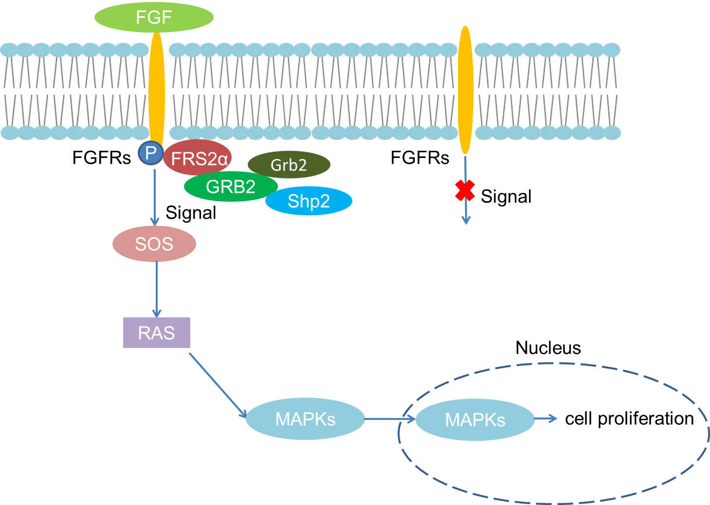 Fig 1. Mechanism of Signaling
Creative Proteomics can provide cytokine detection platform for scientific research. According to different purposes, our dedicated analysts will customize exclusive solutions for you. We aim to provide customers with high-quality and convenient services to help you accelerate the progress of your project.
Our cytokine detection service includes but is not limited to:
- One or more cytokines cytokines qualitative and quantitative detection
- Cytokines qualitative and quantitative detection of various species
- Cytokine antibodies qualitative and quantitative detection
Sample requirements
- Sample Types-Serum, cell supernatant, urine, body fluids, lavage fluid, cerebral spinal cord, atrial fluid, pleural fluid, tissue, etc.
- Sample Volume - It is optimal for at least 200µl of each sample. This volume allows for triplicate testing of each sample.
Our advantages:
- Different detection methods can be selected based on different samples and requirements.
- Ensure the specificity and accuracy of the test by using high quality antibodies.
- Repeat the test to ensure the repeatability and accuracy of the experimental results.
- Feedback results are accurate and efficient
Technology platform:
We mainly provide the Luminex cytokine detection platform. Luminex uses fluorescently encoded microspheres with specific antibodies to different target molecules. The different microspheres can be combined freely to a certain extent so that up to 100 analytes can be tested multiple times simultaneously in a single experiment.
The Luminex cytokine assay platform has the following advantages:
- Multiple detection: simultaneous detection of 100 biological targets
- Short experiment time: 1-3 weeks
- High sensitivity: the lower limit of accurate quantification is as low as 0.1 pg/mL
- Save samples: only need a sample volume as low as 25 μL
- Time saving: the experiment process only takes 4 hours
For your different needs, we can also provide the following detection methods:
- Enzyme-linked immunosorbent assay (ELISA)
- Flow cytometry
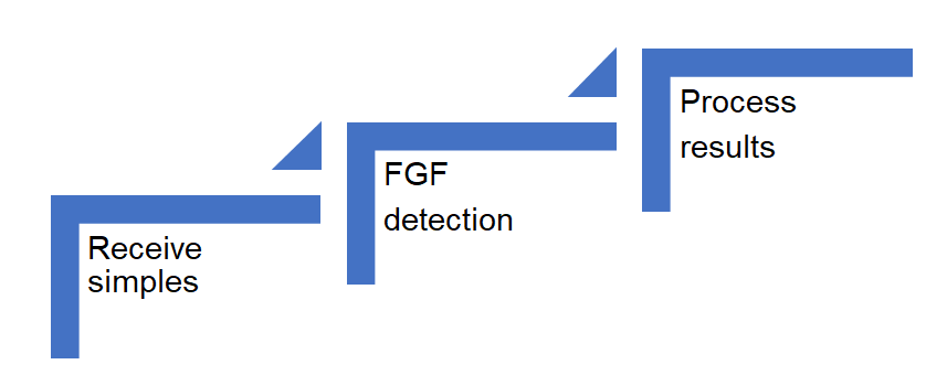
Workflow
For more information about the FGF detection service or need other detection requirements, please contact us.
References:
- Ornitz, D. M., et al. The Fibroblast Growth Factor signaling pathway. Wiley Interdiscip. Rev. Dev. Biol. 4, 215-266.
- Yun, Y.-R., et al. Fibroblast growth factors: biology, function, and application for tissue regeneration. J. Tissue Eng. 2010, 218-142.
- Kouhara H, et al. A lipid-anchored Grb2-binding protein that links FGF-receptor activation to the Ras/MAPK signaling pathway. Cell 1997;89:693–702.