Introduction
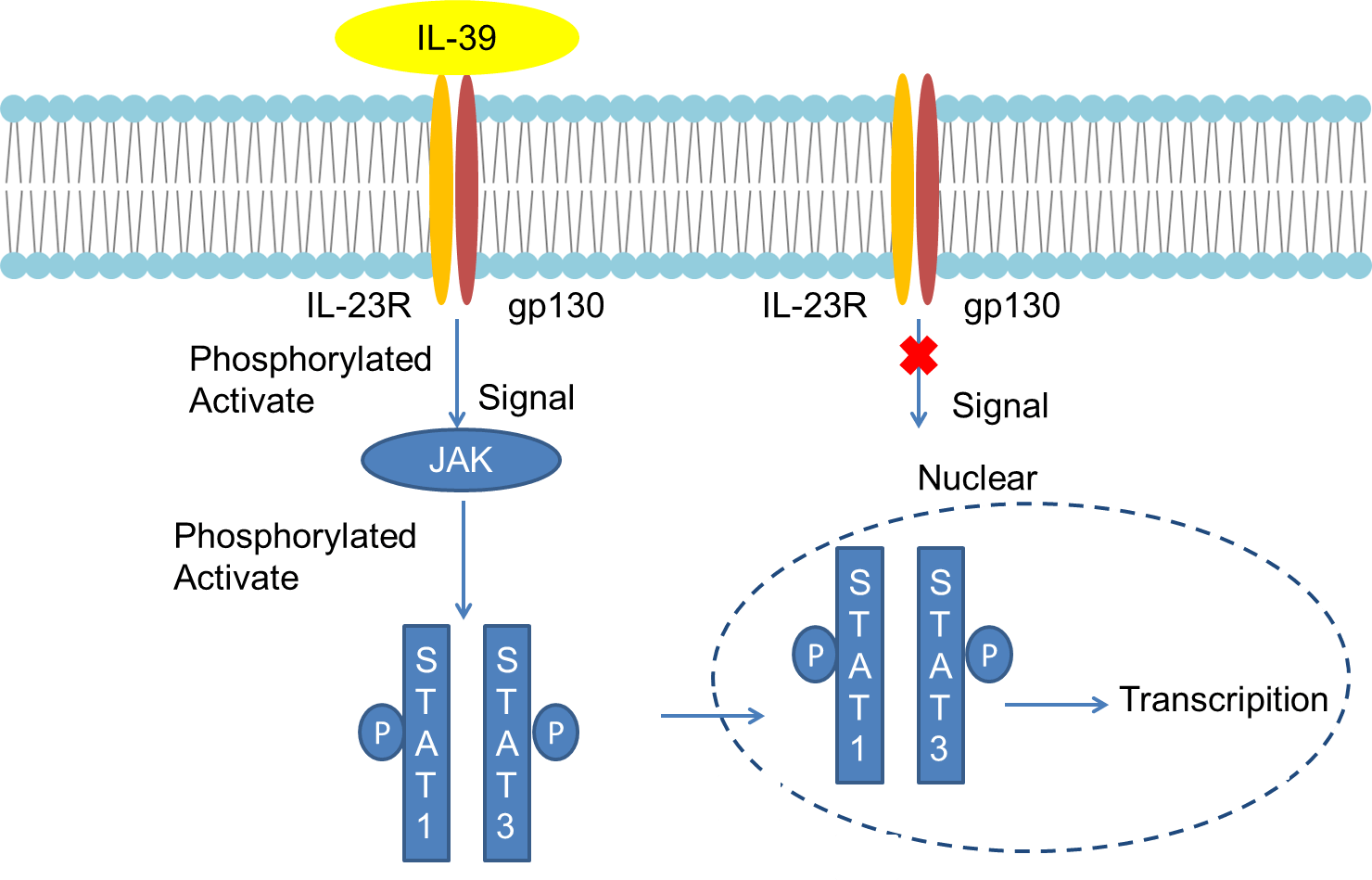
Interleukin-39 (IL-39) is a recently discovered new member of the IL-12 family, consisting of two subunits of IL-23p19 and EB virus-induced gene 3. It is mainly produced by lipopolysaccharide-stimulated B cells and activated GL7 + B cells in lupus mice. IL-23R / gp130 is its receptor, which is activated by signal transduction and transcriptional activation factor 1 (STAT1)/STAT3 signaling pathway mediates inflammation. The current research on IL-39 is still in its infancy. IL-39 may be an important proinflammatory factor, mainly produced by activated B cells in vivo and in vivo, and can be used as a potential therapeutic target for systemic lupus erythematosus.
Mechanism and Function
Cytokines of the interleukin-12 (IL-12) family are heterodimeric glycoproteins composed of alpha and beta polypeptide chains covalently linked by disulfide bonds. The alpha chain includes IL-23p19 , IL27p28 and IL-12p35, and β chain includes IL-12p40 and Epstein-Barr virus-induced gene 3 (EBI3). Six different heterodimers can be formed by combining any one subunit in α chain and β chain. IL-23p19 and EBI3 subunits can combine to form a new heterodimeric protein, and was officially named IL-39. The two subunits of IL-39, IL-23p19 and EBI3, are shared with IL-23 and IL-27 / IL-35, respectively. The IL-23p19 subunit has a 4α helix bundle structure, which has homology with IL-6 and granulocyte colony stimulating factor. The EBI3 subunit was originally found in EB virus-infected B lymphocytes. Its gene is located on human chromosome 17 and encodes a glycoprotein with a relative molecular mass of 34,000, of which 27% have the same amino acid sequence as the IL-12p40 subunit source.
IL-12 family cytokine receptors are composed of homo- or heterodimers formed by IL-12Rβ1, IL-12Rβ2, IL23R, gp130 and IL-27R. Since the subunits of IL-39 are shared with IL-23, IL-27 and IL-35, respectively, it can be reasonably inferred that the receptor of IL-39 is homo- or heterodimerized by IL-23R, IL-27R and gp130. Subsequent experiments proved that IL-39 signaling is carried out through the receptor complex of IL-23R and gp130. The two chains must be aggregated into a receptor complex and bind to IL-39 to activate STAT1 and STAT3 signaling molecules before transmitting signals.
IL-39 is a new member of the IL-12 family (composed of two subunits, IL-23p19 and EBI3). It shares the EBI3 subunit with IL-27 and IL-35. In human B lymphocytes infected with EB virus, EBI3 is high protein expression. At the same time, it was found that after lipopolysaccharides stimulated initial B cells in vitro, both IL-23p19 and EBI3 were expressed and the expression level was positively correlated with the length of lipopolysaccharide stimulation time. In lupus mice, GL7 + B cells and CD138 + plasma cells are highly activated and widely expressed. Activated GL7 + B cells and CD138 + plasma cells can promote high expression of IL-39 in mice, but GL7 + B cells produce IL The protein content of -39 is higher. Studies have shown that there are IL-23p19 and EBI3 mRNA expression in other immune cells such as dendritic cells and macrophages, and its mRNA expression level increases in the presence of IL-4. However, the study found that lipopolysaccharide has an inhibitory effect on the mRNA expression of IL-23p19 and EBI3 in dendritic cells and macrophages. These results indicate that there is a complex mechanism for the expression of IL-23p19 and EBI3 in lipopolysaccharide and dendritic cells, macrophages, and B cells, and further research is needed.
 Fig 1. Mechanism of Signaling
Creative Proteomics can provide cytokine detection platform for scientific research. According to different purposes, our dedicated analysts will customize exclusive solutions for you. We aim to provide customers with high-quality and convenient services to help you accelerate the progress of your project.
Our cytokine detection service includes but is not limited to:
- One or more cytokines cytokines qualitative and quantitative detection
- Cytokines qualitative and quantitative detection of various species
- Cytokine antibodies qualitative and quantitative detection
Sample requirements
- Sample Types-Blood, serum, plasma, cell culture medium, tissue homogenate, etc.
- Sample Volume - It is optimal for 50 samples. This volume allows for triplicate testing of each sample.
Our advantages:
- Different detection methods can be selected based on different samples and requirements.
- Ensure the specificity and accuracy of the test by using high quality antibodies.
- Repeat the test to ensure the repeatability and accuracy of the experimental results.
- Feedback results are accurate and efficient
Technology platform:
We mainly provide the Luminex cytokine detection platform. Luminex uses fluorescently encoded microspheres with specific antibodies to different target molecules. The different microspheres can be combined freely to a certain extent so that up to 100 analytes can be tested multiple times simultaneously in a single experiment.
The Luminex cytokine assay platform has the following advantages:
- Multiple detection: simultaneous detection of 100 biological targets
- Short experiment time: 1-3 weeks
- High sensitivity: the lower limit of accurate quantification is as low as 0.1 pg/mL
- Save samples: only need a sample volume as low as 25 μL
- Time saving: the experiment process only takes 4 hours
For your different needs, we can also provide the following detection methods:
- Enzyme-linked immunosorbent assay (ELISA)
- Flow cytometry
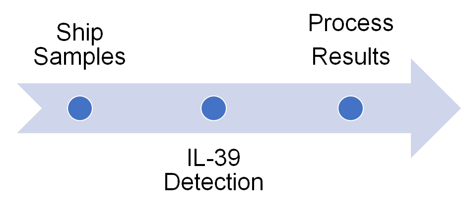
Workflow

For more information about the IL-39 detection service or need other detection requirements, please contact us.
References:
- Wang X, et al. A novel IL-23p19/Ebi3 (IL-39) cytokine mediates inflammation in Lupuslike mice. Eur J Immunol, 2016, Mar 28. doi: 10.1002/ eji.201546095
- Ringkowski S, et al. Interleukin-12 family cytokines and sarcoidosis. Front Pharmacol, 2014, 5: 233

