
Introduction
IL-3 is a hematopoietic growth factor and is mainly produced by activated T cells. It can promote the proliferation of early hematopoietic progenitor cells. Besides, IL-3 can also promote the differentiation of hematopoietic progenitor cells together with erythropoietin granulocyte colony-stimulating factor(G-CFS), and macrophage colony-stimulating factor(M-CFS). It is important for the body's hematopoietic and immune regulatory functions. In addition, IL-3 can also participate in the development of the central nervous system, promote the proliferation and survival of neural precursor cells, then affect brain capacity. In the clinic, the combination of IL-3 with GM-CFS, G-CSF, M-CSF, or EPO after chemotherapy can appropriately stimulate the production of blood cells and platelets, which can rebuild bone marrow function when bone marrow suppression caused by chemotherapy and radiotherapy. Besides, the combination can also improve aplastic anemia. In diseases, IL-3 can also regulate eosinophils and other inflammatory cells, and it is the key factor to start and sustain asthma airway inflammation.
Mechanism and Function
The IL-3 receptor is a heterodimer composed of two chains of α and β, with a molecular weight of 140 kD. The α chain is unique to IL-3, and also determines the specificity of IL-3 action. The β chain is shared with GM-CSF and IL-5. Among them, only a single α chain can form a high affinity receptor. The signal transduction of IL-3 in the regulation of hematopoietic stem cell proliferation and differentiation is mainly achieved through the JAK2 and Src kinases. When IL-3 functions, IL-3 first forms heterodimers or oligomers. After the oligomers polymerize, it stimulates intracellular signal transduction. Among the α and β chains of the IL-3 receptor, the α chain has no signal transduction function, and intracellular signals are mainly transmitted by the β chain. The IL-3R cytoplasm interacts with the Scr family tyrosine kinases such as Lyn and Fyn, thereby activating the ras-MAPK pathway. MAPK can activate STAT and act on promoters of related genes, thereby regulating related gene expression.
After IL-3R forms a hetero oligomer, IL-3 can bring the near-membrane part of the receptor closer to JAK2. JAK2 binds to the β chain, causing further aggregation of JAK2. Corresponding STATs were then recruited in the cytoplasm. STATs form complexes with activated receptor polymers and are then phosphorylated. Dimerized STAT escapes from the complex, passes through the nucleus and enters the nucleus, initiates the transcription of specific genes, then transcribes hematopoietic stem cells. Through the above mechanism, IL-3 participates in.
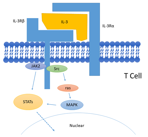 Fig 1. Mechanism of Signaling
Creative Proteomics can provide cytokine detection platform for scientific research. According to different purposes, our dedicated analysts will customize exclusive solutions for you. We aim to provide customers with high-quality and convenient services to help you accelerate the progress of your project.
Our cytokine detection service includes but is not limited to:
- Quantitative and qualitative detection of cytokines in different species
- Quantitative and qualitative detection of cytokine antibodies
- Quantitative and qualitative detection of single/mulltiple cytokines
Sample requirements
- Sample Types: Blood, serum, plasma, cerebrospinal fluid, cell culture supernatant, tissue homogenate, cell culture medium, urine, tumor, etc.
- Sample Volume: It is optimal for at least 200µl of each sample. This volume allows for triplicate testing of each sample.
Our advantages:
- Efficient design: Multiplex reaction detecting in various methods are available, which can be selected according to different samples and requirements.
- Sensitive detection: High-quality antibodies are used in the detection process to improve detection specificity and accuracy.
- Reliable results: The feedback results are professional and efficient.
Technology platform:
We mainly provide the Luminex cytokine detection platform. Luminex uses fluorescently encoded microspheres with specific antibodies to different target molecules. The different microspheres can be combined freely to a certain extent so that up to 100 analytes can be tested multiple times simultaneously in a single experiment.
The Luminex cytokine assay platform has the following advantages:
- Multiple detection: simultaneous detection of 100 biological targets
- Short experiment time: 1-3 weeks
- High sensitivity: the lower limit of accurate quantification is as low as 0.1 pg/mL
- Save samples: only need a sample volume as low as 25 μL
- Time saving: the experiment process only takes 4 hours
For your different needs, we can also provide the following detection methods:
- Enzyme-linked immunosorbent assay (ELISA)
- Flow cytometry
Workflow
 Fig 2. workflow
For more information about the IL-3 detection service or need other detection requirements, please contact us.
References:
- Reddy E P, et al. IL-3 signaling and the role of Src kinases, JAKs and STATs: a covert liaison unveiled. Oncogene, 2000, 19(21):2532-2547.
- Carr P D, et al. Structure of the complete extracellular domain of the common beta subunit of the human GM-CSF, IL-3, and IL-5 receptors reveals a novel dimer configuration. Cell, 2001, 104(2):291-300.