
Introduction
Creative Proteomics can simultaneously quantitatively detect atopic dermatitis-related cytokines and chemokines through a variety of technologies, and provide higher sensitivity and a wider detection range.
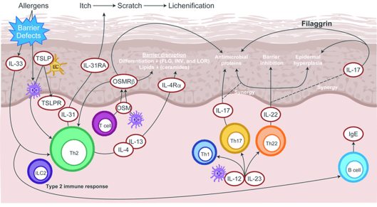
Atopic dermatitis (AD) is a skin disease that is genetically prone to chronic recurrence and high itching caused by the destruction of the epidermal barrier and related immune disorders. Most AD is onset in early childhood and has characteristics that depend on age distribution. The disease is usually accompanied by elevated IgE levels, increased peripheral blood eosinophils, and other allergic symptoms.
T Lymphocyte subsets and cytokines in AD
In the acute phase of AD, Th2 differentiation is dominant in the presence of IL-4. Th2 response leads to an increase in the production of cytokines, mainly including IL-4, IL-5, IL-13, IL-31 and other cytokines, and promotes the pathogenesis of AD.
Naive T lymphocytes are induced to differentiate into Th 22 cells in an IL-6 and TNF-dependent manner. The main transcription factor of human Th 22 differentiation is aryl hydrocarbon receptor. Receptor binding is activated by STAT 3 to produce cytokines such as IL-22, IL-26 and IL-13.
Under the combined action of TGF-β and IL-4, the initial CD4+ T lymphocytes can be directly transformed into Th 9 cells. Co-stimulatory molecules such as CD28 and OX40 induce an increase in Th 9 production under the synergistic effect of TGF-β and IL-4. However, IFN-γ and IFN-γ-promoting cytokines such as IL-12, IL-18, and IL-23, and Th 1 related transcription factor T-bet inhibit the induction of Th 9 cells. Th 9 cells selectively produce IL-9 and also secrete IL-10 and IL-21.
IL-6 and TGF-β1 together induce Th 17 differentiation. IL-21 produced by Th 17 cells can also promote Th 17 differentiation through autocrine. IL-23 contributes to the survival and proliferation of previously differentiated Th17 cells, and can enhance the pathogenicity of Th17 cells in the presence of IL-6. Th 17 functions mainly by secreting cytokines, mainly including IL-17A (IL-17), IL-17F, IL-21, IL-22, GSF and potentially TNF-α, IL-6 and IL-8, to promote the production of acute inflammation.
In addition to the above cytokines, IL-18, IL-33, and TSLP also play an important role in the inflammatory response of AD skin lesions.
Detectable cytokines include but are not limited to:
| IL-4 | IL-5 | IL-13 | IL-31 | IL-6 |
| IL-22 | IL-26 | IL-13 | IL-12 | IL-18 |
| IL-23 | IL-10 | IL-21 | TGF-β | IFN-γ |
| TNF-α |
Technology platform
We mainly provide the Luminex cytokine detection platform. Luminex uses fluorescently encoded microspheres with specific antibodies to different target molecules. The different microspheres can be combined freely to a certain extent so that up to 100 analytes can be tested multiple times simultaneously in a single experiment.
The Luminex cytokine assay platform has the following advantages:
- Multiple detection: simultaneous detection of 100 biological targets
- Short experiment time: 1-3 weeks
- High sensitivity: the lower limit of accurate quantification is as low as 0.1 pg/mL
- Save samples: only need a sample volume as low as 25 μL
- Time saving: the experiment process only takes 4 hours
For your different needs, we can also provide the following detection methods:
- Flow cytometry (FACS analysis): Identify and measure cell biomarkers in complex subpopulations. It can be used for intracellular cytokine detection without two hours.
- Enzyme-linked immunosorbent assay (ELISA): Use the primary antibody for capture, and conjugate the secondary antibody with an enzyme or radioisotope for detection. Our multiplexing system can detect the expression of multiple cytokines at once.
Sample preparation
- Suitable for serum, plasma, cell culture supernatant and all kinds of body fluids.
- The body fluid samples are stored in a refrigerator at -80 degrees Celsius and transported on dry ice.
Creative Proteomics can provide you with a one-stop solution. If you want to test other cytokines or other information you want to consult, please contact us. We are looking forward to cooperating with you.
Reference:
- Smith P, Howell M, Kuo F. Targeting the Janus kinase family in autoimmune skin diseases. Frontiers in immunology, 2019, 10: 2342.