
The guinea pig occupies a distinctive niche in immunological research. It is the gold-standard small-animal model for tuberculosis vaccine studies because it develops caseating granulomas — the hallmark of human TB pathology — that are absent in mouse models. It is the primary model for RSV and CMV pathogenesis because its lung physiology closely mirrors the human lung. It is widely used in regulatory inhaled toxicology and contact hypersensitivity research because its skin and pulmonary immune responses translate more directly to humans.
Yet the guinea pig has the poorest commercial Luminex multiplex kit coverage of any major research species. Human and mouse researchers choose from dozens of pre-configured 20- to 40-plex panels; guinea pig researchers have a single 5-plex commercial kit. This gap means that virtually every guinea pig cytokine study — whether a 10-animal pilot or a 110-animal vaccine efficacy trial — requires a custom-designed panel built from individually validated antibody pairs.
Why the Guinea Pig Model Matters for Immunology Research
- Tuberculosis vaccine research: Guinea pigs develop caseating granulomas indistinguishable from those in human TB patients. Mouse models do not recapitulate this pathology. Vaccine efficacy studies require tracking Th1/Th2 cytokine balance (IFN-γ, IL-2, IL-4, IL-10, TNF-α) across pre-vaccination, post-vaccination, post-challenge, and post-necropsy timepoints (Clark et al., 2015; Padilla-Carlin et al., 2008).
- Respiratory viral pathogenesis: The guinea pig lung mirrors the human lung in airway branching, mucus-producing goblet cell distribution, and cough reflex — features that make it the standard model for RSV and CMV studies.
- Inhaled toxicology: Regulatory toxicology guidelines frequently specify guinea pig inhalation exposure studies; cytokine profiling in BALF is the primary readout for pulmonary inflammation.
- Contact hypersensitivity and skin immunology: Guinea pig Langerhans cell distribution and DTH kinetics more closely mirror human skin than mouse models, making it the preferred species for sensitisation and adjuvant safety testing.
Guinea Pig Luminex Cytokine Panels: What Can Be Measured
Approximately 40 analytes have validated antibody pairs for guinea pig Luminex detection — more than most researchers assume — but every panel must be assembled de novo. A single commercial 5-plex (IL-1β, IL-6, IL-8, MIP-1β, TNF-α) exists as a starting point; for any study requiring 6+ analytes, a custom panel is the only option.
| Category | Available Analytes |
|---|---|
| Interleukins | IL-1β, IL-2, IL-4, IL-6, IL-10, IL-11, IL-12, IL-13, IL-15, IL-16, IL-17A, IL-18, IL-21, IL-22, IL-23, IL-27, IL-33 |
| TNF family & interferons | TNF-α, TNF-β, IFN-γ |
| Chemokines | CCL2, CCL5 (RANTES), CCL20, CXCL8 (IL-8), CXCL10 (IP-10), Eotaxin, MCP-1, MIP-1α, MIP-1β |
| Growth factors | GM-CSF, VEGF, PDGF, FGF-basic, EGF, IGF-1, BMP-2, NGF, SCF, G-CSF, LIF, TGF-β |
For panel configuration, use our Luminex Multiplex Assay Customization Tool. For studies requiring species-specific guinea pig validation, our Guinea Pig Cytokine Assay service provides dedicated support.
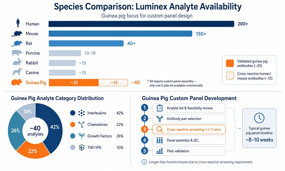 Guinea pig Luminex analyte landscape: species comparison showing that guinea pig has 40+ validated targets but no pre-configured panels, with category distribution and custom development timeline including cross-reactive antibody screening.
Cross-Reactive Antibody Validation for Guinea Pig Panels
Because the guinea pig antibody catalogue is limited, a substantial proportion of custom panels rely on cross-reactive antibodies — those raised against human or mouse targets that also recognise the orthologous guinea pig protein. This can expand a panel by 30–50% but requires additional validation:
- Sequence homology screening: Guinea pig protein sequence is aligned with human/mouse orthologues. Regions with ≥80% identity in the antibody epitope region are candidates for cross-reactivity.
- Binding verification: The candidate antibody pair is tested against recombinant guinea pig protein or endogenous protein in guinea pig matrix before entering the multiplex cross-reactivity matrix.
- Multiplex cross-reactivity testing: The pair enters the full N×N matrix with other panel members. The standard <1% cross-signal threshold applies.
- Matrix parallelism: Dilution linearity is tested in the target guinea pig matrix to confirm proportional performance.
Cross-reactive antibodies are not a shortcut — they add 2–3 weeks per analyte to the validation timeline but are often essential for achieving a scientifically meaningful panel for this species.
Designing a Large-Cohort Guinea Pig Cytokine Study
The most common design: 50–110 animals, 3–4 treatment groups, 3–6 timepoints. A 110-animal, 4-group, 3-timepoint study generates ~330 samples; an 8-plex panel translates to ~350 Luminex wells across 4–5 plates.
Sample volume planning
- Terminal bleed (cardiac puncture): 2–3 mL whole blood → ~1–1.5 mL serum or plasma.
- Non-terminal bleed (saphenous/jugular): 0.5–1 mL whole blood → ~250–500 µL serum or plasma.
- BALF: 1–2 mL per animal, collected at necropsy.
Luminex requires 25 µL per well regardless of plex size. At 8-plex, the same 25 µL measures all eight analytes. Terminal bleeds support all timepoints; non-terminal bleeds support one timepoint per bleed. Cumulative non-terminal volume should not exceed 10% of total blood volume (~25 mL for a 400 g animal).
Plate design and QC
- Pooled QC sample: Pool from 8–12 representative samples. Run in duplicate on every plate. Inter-plate %CV ≤20%.
- Bridging controls: 3–4 samples on every plate to anchor cross-plate comparability.
- Randomisation: Randomise animals across plates, not by treatment group.
- Blind QC samples: 1–2 blinded duplicate pairs per plate — the same sample under different IDs in non-adjacent wells. Discordant blind duplicates indicate plate-level handling errors that automated QC cannot catch.
- Drift monitoring: 7-point standard + blank at start and end of each plate.
Reporting values below LLOQ
- Raw reporting: Values below LLOQ are reported as "<LLOQ" with the plate-specific LLOQ noted. Standard for regulatory-facing studies.
- Imputation: If statistical modelling requires numerical values, the report includes both raw and imputed datasets. Imputation method must be pre-specified in the statistical analysis plan.
- Analyte exclusion: If >50% of samples in all groups return <LLOQ, that analyte is excluded from formal testing and reported descriptively only.
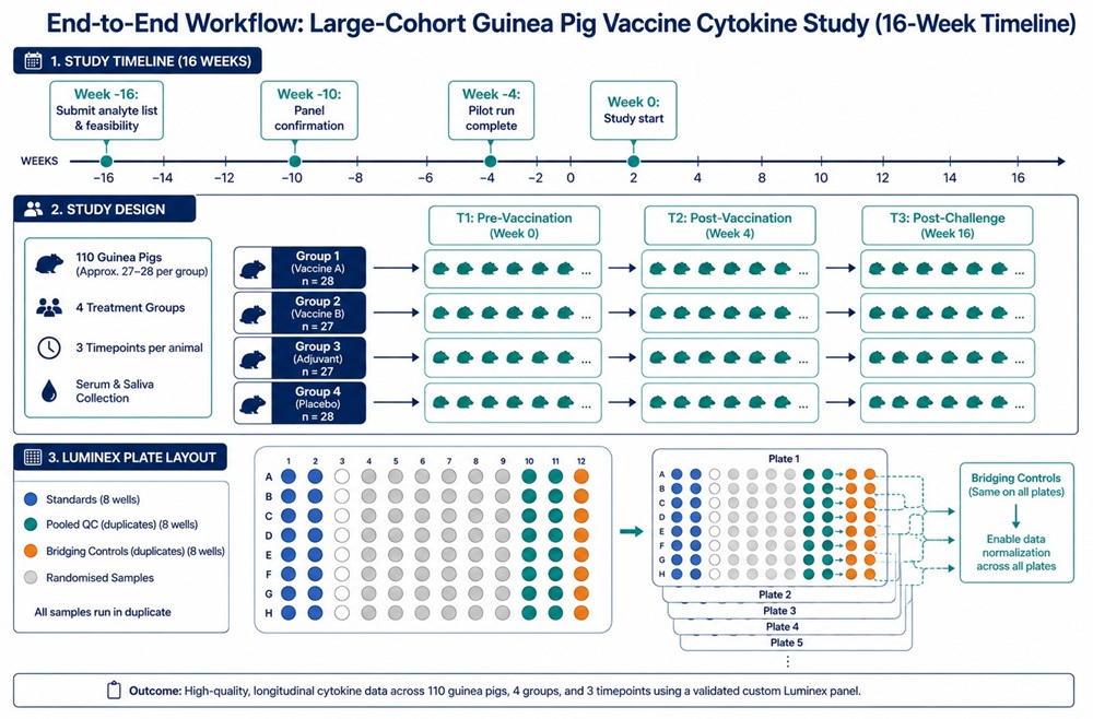 End-to-end workflow for a 110-animal guinea pig vaccine cytokine study: panel development timeline, study design schematic with four treatment groups and three timepoints, and randomised multi-plate QC layout with bridging controls.
Guinea Pig Sample Collection: Blood, BALF, and Tissue Requirements
- Serum (preferred): Serum separator tubes; clot at RT 30 min; centrifuge 1,500–2,000 × g, 10 min, 4°C. Aliquot into single-use 75–100 µL volumes.
- EDTA-plasma: Acceptable for most analytes. Specify anticoagulant when requesting panel validation.
- BALF: Centrifuge 300–400 × g, 10 min, 4°C to remove cells. BALF cytokine concentrations are 5–10× lower than paired serum — factor this into LLOQ expectations.
- Tissue homogenate: Homogenise at 100 mg/mL in PBS with protease inhibitors; centrifuge 10,000 × g, 15 min; normalise total protein across samples.
Storage and shipping
- Single-use aliquots labelled with ID, date, and freeze-thaw count.
- Snap-freeze immediately after processing.
- Store at −80°C. IL-6 and TNF-α degrade after 2 freeze-thaw cycles.
- Ship on dry ice with temperature loggers; do not use wet ice.
International shipping logistics
- Dry ice provisioning: 200–400 cryovials require ~10–15 kg dry ice (domestic overnight) or 20–25 kg (international 2–3 day). Dry ice sublimates at ~2–4 kg per 24 hours.
- Temperature monitoring: Calibrated data logger inside the sample box, alarming if temperature rises above −60°C (dry ice sublimation point is −78.5°C).
- Customs: Commercial invoice describing contents as "non-infectious research samples — animal origin". Import permits may be required by destination country.
- Timing: Ship Monday–Wednesday only for international destinations; weekend customs delays can exhaust dry ice.
- Receipt verification: Receiving lab checks data logger, confirms samples are frozen, photographs the opened container, and transfers immediately to −80°C.
Panel Configuration Decisions: One Panel or Two?
- Acceptable cross-reactivity (<1%): All analytes pass. Single multiplex assembled.
- Borderline (1–5%): If the interferent is 50–100× lower concentration than the affected target, accept with documented caveat. Otherwise, replace antibody or split.
- Unacceptable (>5%): Split into separate panels. Up to 2–3 replacement antibody rounds tested before splitting.
Split-panel designs approximately double sample consumption (~75 µL → ~150 µL per timepoint) but fully preserve data quality. Splitting is not a failure — it is a standard resolution, and more common in guinea pig work than human or mouse.
Custom Panel Development Timeline for Guinea Pig Studies
| Phase | Timeline |
|---|---|
| Analyte selection & feasibility | 1–2 weeks |
| Cross-reactive antibody validation (if needed) | +2–3 weeks |
| Cross-reactivity testing | 3–4 weeks |
| Standard curve validation | 2–3 weeks |
| Matrix validation (spike-recovery, dilution linearity) | 2 weeks |
| Pilot run | 1–2 weeks |
| Total (standard) | 9–13 weeks |
| Total (with cross-reactive screening) | 11–16 weeks |
Planning Your Guinea Pig Cytokine Study: Timeline and Logistics
- 3–4 months before first sample collection: Submit analyte list and matrix for feasibility assessment. Begin cross-reactive antibody screening if needed.
- 8–10 weeks before collection: Confirm panel composition after cross-reactivity testing. Begin validation.
- 2–4 weeks before collection: Complete pilot run on representative samples.
- Study start: Samples flow directly into a validated, pilot-tested assay pipeline.
For consultation on panel design, see our Luminex cytokine detection service.
References:
- Clark, Simon, et al. "Animal models of tuberculosis: guinea pigs." Cold Spring Harbor Perspectives in Medicine 5.5 (2015): a018549. DOI: 10.1101/cshperspect.a018549
- Padilla-Carlin, Danielle J., et al. "The guinea pig as a model of infectious diseases." Comparative Medicine 58.4 (2008): 324–340. PMID: 18724769
- Orme, Ian M., and Diane J. Ordway. "Host response to nontuberculous mycobacterial infections of current clinical importance." Infection and Immunity 82.9 (2014): 3516–3522. DOI: 10.1128/IAI.01906-14
- Vignali, D. A. A. "Multiplexed particle-based flow cytometric assays." Journal of Immunological Methods 243.1–2 (2000): 243–255. DOI: 10.1016/S0022-1759(00)00238-6
- Tighe, P. J., et al. "ELISA in the multiplex era: Potentials and pitfalls." Proteomics – Clinical Applications 9.3–4 (2015): 406–422. DOI: 10.1002/prca.201400130