Comprehensive multiplex panels for immuno-oncology biomarker discovery — 111 to 131 analytes spanning soluble immune checkpoint proteins, cytokines, chemokines, cytotoxic effector molecules, angiogenic factors, and matrix remodeling enzymes in a single 25 µL sample.
Immuno-oncology has transformed cancer treatment, but predicting and monitoring therapeutic response requires moving beyond single-marker approaches. The tumor microenvironment is a complex network of immune checkpoint signaling (PD-1/PD-L1, CTLA-4, LAG3, TIM-3, TIGIT), T-cell effector function (Granzyme B, Perforin), cytokine-mediated immune polarization, chemokine-driven cell trafficking, and angiogenic remodeling — all interacting simultaneously. Our MSD Immuno-Oncology panels capture this multi-dimensional biology in one assay, providing the breadth needed to characterize the TME, monitor pharmacodynamic responses to immunotherapy, and discover composite biomarker signatures that no single analyte can reveal.
Cancer immunotherapy — checkpoint inhibitors, bispecific antibodies, CAR-T cells, cancer vaccines — all converge on a common mechanism: modulating the immune system's ability to recognize and eliminate tumor cells. But response is heterogeneous. Some patients achieve durable remission; others progress. Identifying which patients respond, and why, requires profiling the full network of immune signals — not just one checkpoint or one cytokine.
Soluble immune checkpoint proteins shed into circulation reflect the expression and engagement of their membrane-bound counterparts in the TME. When PD-L1 is expressed on tumor cells, its soluble form (sPD-L1) increases in plasma. When T cells upregulate LAG3 or TIM-3 as exhaustion markers, their soluble forms become detectable. Measuring 15+ soluble checkpoints simultaneously — alongside cytokines reflecting T-cell polarization, chemokines directing immune infiltration, and effector molecules (Granzyme B, Perforin) reporting cytotoxic activity — provides a systems-level view of the anti-tumor immune response that no single analyte can deliver.
Two pre-configured configurations for Immuno-Oncology biomarker discovery, differentiated by the inclusion of cytotoxic effector molecules and tumor-stroma interface markers.
| Panel | Plex | Core Analyte Categories | Unique Coverage | Species |
|---|---|---|---|---|
| MSD Immuno-Oncology 111-Plex | 111 | Immune checkpoints, cytokines, chemokines, growth factors, metabolic hormones | Core checkpoint + cytokine profiling for Immuno-Oncology biomarker programs | Human |
| MSD Immuno-Oncology 131-Plex | 131 | 111-Plex + cytotoxic effectors + tumor-stroma interface markers | Granzyme A/B, Perforin, MMP-1/2/7/9, Galectin-9, HVEM, Nectin-4, Pentraxin 3, S100A12, E-Selectin, P-Selectin, RAGE, CD27, LIGHT, APRIL | Human |
| MSD U-PLEX Custom Immuno-Oncology Panel | 2–10 | Select specific checkpoint + cytokine targets from U-PLEX catalog | Focused validation panels from 111/131-Plex discovery hits | Human |
The 111-Plex provides comprehensive checkpoint + cytokine coverage. The 131-Plex adds 20 cytotoxic effector, matrix remodeling, and tumor-stroma interface markers for deeper TME characterization. Panel selection depends on whether T-cell cytotoxic function and stromal remodeling are central to your research question. All services are for research use only.
The 111-Plex and 131-Plex include the most comprehensive soluble immune checkpoint coverage available in a single multiplex panel — spanning targets of approved immunotherapies, investigational agents, and co-stimulatory receptors.
PD-1 (epitope 1 & 2), PD-L1/B7-H1 (epitope 1 & 2), PD-L2, CTLA-4, LAG3, HAVCR2/TIM-3, TIGIT — the inhibitory checkpoint receptors and ligands targeted by approved and late-stage investigational immunotherapies. Dual-epitope PD-1 and PD-L1 assays distinguish different molecular forms and drug-bound vs. free receptor.
CD28, ICOS, OX40/TNFRSF4, GITR/TNFRSF18, CD27, HVEM/TNFRSF14, CD40L (soluble) — the co-stimulatory receptors that amplify T-cell responses and are targets of agonist immunotherapies (GITR, OX40, CD27, ICOS agonists in clinical development).
BAFF, BAFF-R/TNFRSF13C, BCMA/TNFRSF17, APRIL/TNFSF13, CD20 — B-cell lineage markers and survival factors. Relevant for CAR-T target antigen monitoring (BCMA for multiple myeloma CAR-T), B-cell depletion therapy pharmacodynamics (CD20), and tertiary lymphoid structure (TLS) research in the TME.
Granzyme A, Granzyme B, Perforin — the three primary effector molecules of cytotoxic T lymphocytes and NK cells. Direct readout of immune-mediated tumor cell killing. Elevated in responders to checkpoint inhibitor therapy; suppressed in T-cell exhaustion. Exclusive to the 131-Plex configuration.
MMP-1, MMP-2, MMP-7, MMP-9, Galectin-9, Nectin-4, E-Selectin, P-Selectin — enzymes and adhesion molecules mediating extracellular matrix remodeling, immune cell extravasation, and tumor-stroma cross-talk. Reflect the architectural changes in the TME during immune-mediated tumor rejection.
Pentraxin 3, S100A12, RAGE/AGER, LIGHT/TNFSF14, APRIL/TNFSF13, GITRL/TNFSF18, ICOS-L/B7-H2 — soluble mediators amplifying innate and adaptive immune responses in the TME. Reflect the inflammatory context in which checkpoint blockade operates.
Consult on your Immuno-Oncology research objectives. 111-Plex for checkpoint + cytokine coverage; 131-Plex to add cytotoxic effectors and stromal markers. We advise on sample size considerations for high-dimensional biomarker discovery.
EDTA plasma, processed within 1 hour of collection, frozen at −80°C. For longitudinal Immuno-Oncologystudies, consistent time-of-day collection and pre-treatment vs. on-treatment timepoint documentation is essential.
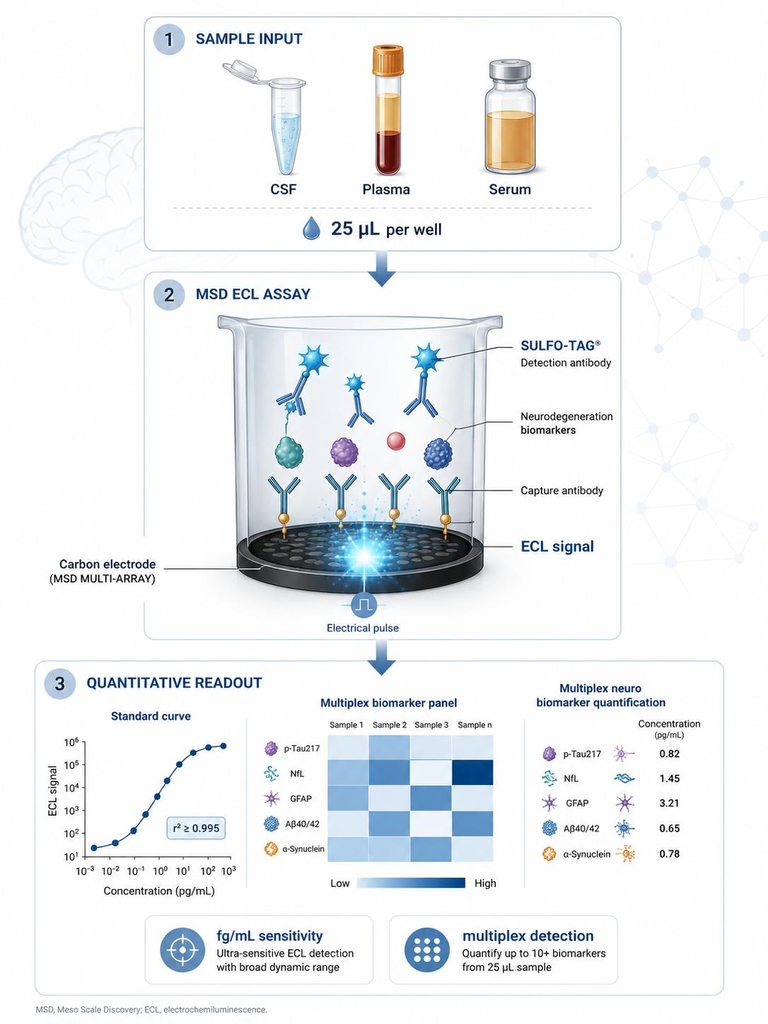
111 or 131 analytes measured simultaneously from 25 µL. Multi-analyte 8-point standard curves, three-level QC controls, blank wells. ~70 seconds read time per plate.
Comprehensive report: concentrations for all analytes organized by functional category (checkpoints, cytokines, chemokines, effectors, stromal), QC metrics, raw ECL data, and methods summary.
Primary matrix for soluble checkpoint and cytokine panels. Collect in EDTA tubes, centrifuge within 1 hour at 1,500×g for 10 min at 4°C. Transfer to polypropylene cryovials, freeze at −80°C. Consistent collection time relative to treatment cycle is essential.
Acceptable alternative for most panel analytes. SST collection, 30 min clotting at RT, centrifuge at 1,500×g for 10 min at 4°C. Note: clotting releases platelet-derived factors; maintain consistent matrix within a study.
For CNS tumors, brain metastasis, and ICANS biomarker studies. Collect in polypropylene tubes, centrifuge at 2,000×g for 10 min at 4°C, aliquot, freeze at −80°C. Compatible with limited volumes (25 µL per panel).
For direct TME biomarker measurement at the tumor site. Flash-freeze biopsies, homogenize in RIPA buffer with protease/phosphatase inhibitors, centrifuge at 14,000×g for 15 min at 4°C. Normalize to total protein. Captures local checkpoint, cytokine, and MMP concentrations that may differ substantially from systemic levels.
For in vitro IO models: T-cell:tumor co-cultures, CAR-T cytotoxicity assays, macrophage polarization, organoid-immune co-cultures. Centrifuge at 300×g for 5 min, aliquot, freeze at −80°C. 25 µL enables serial sampling from the same culture well.
Malignant effusions are a direct window into the local TME in advanced cancer patients. Centrifuge at 1,500×g for 10 min to remove cells and debris, aliquot, freeze at −80°C. High protein content may require dilution optimization — contact us for guidance.
Longitudinal sampling: pre-treatment baseline, on-treatment (typically Cycle 2–3 Day 1 pre-dose), and at progression or response assessment. Document treatment regimen, cycle number, days since last dose, concomitant corticosteroid use, and irAE status for each timepoint. Shipping: dry ice, overnight courier. Most cytokines and soluble checkpoints stable ≥12 months at −80°C with ≤2 freeze-thaw cycles. Single-use aliquots recommended. Repeated freeze-thaw degrades complement factors and MMPs — relevant for 131-Plex stromal markers.
Concentrations for all 111 or 131 analytes organized by functional category (checkpoints, cytokines, chemokines, effectors, stromal). QC metrics per analyte. Excel + PDF.
Raw ECL signal values, per-analyte standard curve plots, QC trend charts, plate layout maps. Compatible with R, Python, Prism.
Detailed protocol including panel configuration, assay conditions, and QC criteria — formatted for manuscript Methods sections in Immuno-Oncology research publications.
The choice depends on whether T-cell cytotoxic function and stromal remodeling are central to your research question. 111-Plex: comprehensive checkpoint + cytokine + chemokine coverage — sufficient for most Immuno-Oncology biomarker programs focused on immune checkpoint biology and cytokine-mediated immune polarization. 131-Plex: adds Granzyme A, Granzyme B, Perforin (direct T-cell/NK cytotoxic readout), MMP-1/2/7/9, E-Selectin, P-Selectin (stromal remodeling), Galectin-9, HVEM, Nectin-4, Pentraxin 3, S100A12, RAGE, CD27, LIGHT, APRIL (tumor-stroma interface and inflammatory amplification). If your research involves tracking T-cell cytotoxic activity during immunotherapy, evaluating stromal barriers to immune infiltration, or studying MMP-mediated checkpoint shedding, the 131-Plex is the appropriate choice. Our scientific team can advise during study design.
Soluble and membrane-bound checkpoint measurements answer different questions. Flow cytometry tells you which cell types express checkpoint proteins and at what level — it answers "which cells?" MSD multiplex quantifies the shed/soluble forms in circulation — it answers "how much total checkpoint signal?" The two are complementary: soluble PD-L1 levels reflect overall tumor and immune cell PD-L1 expression burden; soluble LAG3 and TIM-3 reflect the degree of systemic T-cell exhaustion. Soluble checkpoints have the additional advantage of being measurable from frozen plasma — enabling retrospective analysis of banked research study samples where viable cells for flow cytometry are unavailable. Many Immuno-Oncology research programs use both: flow cytometry for cellular phenotyping and MSD for systemic soluble checkpoint quantification.
Yes. The 111-Plex and 131-Plex include the targets of approved checkpoint inhibitors (PD-1, PD-L1, CTLA-4, LAG3) and numerous investigational targets. Longitudinal measurement of soluble checkpoint and cytokine levels during immunotherapy can reveal pharmacodynamic patterns: increases in Granzyme B and Perforin (131-Plex) suggest T-cell activation; shifts in cytokine profiles from immunosuppressive (IL-10, TGF-β-associated) to pro-inflammatory (IFN-γ, TNF-α, IL-2) suggest immune polarization; decreases in soluble PD-L1 may reflect reduced tumor burden. All applications are for research use only — these panels use manufacturer-documented MSD reagents.
Key recommendations for Immuno-Oncology clinical research sample preparation: (1) Standardize collection time relative to dosing — pre-dose (trough) samples are generally preferred for pharmacodynamic biomarker consistency; (2) Document concomitant medications — particularly corticosteroids (which suppress multiple cytokines) and growth factors (G-CSF, which affects myeloid markers); (3) Record irAE status at each timepoint — immune-related adverse events produce acute-phase cytokine changes that should be distinguished from anti-tumor immune activation; (4) Use consistent tube type and processing protocol throughout the study — EDTA plasma, processing within 1 hour, single-use aliquots at −80°C. Contact us for detailed sample collection SOPs tailored to your trial design.
Online Inquiry
×