Comprehensive multiplex cytokine, chemokine, and immune mediator profiling powered by Electrochemiluminescence (ECL). From focused 4-plex inflammation panels to the flagship 54-plex biomarker discovery platform — V-PLEX, U-PLEX, and S-PLEX across human, mouse, rat, and NHP.
Cytokines rarely act in isolation — they function in coordinated networks where pro-inflammatory, anti-inflammatory, and chemotactic signals intersect to govern immune responses. MSD's spatially separated ECL detection enables simultaneous quantification of up to 54 analytes from a single 25 µL sample, without the spectral overlap, sensitivity compromises, or matrix interference that limit bead-based multiplex platforms. For preclinical research, translational biomarker programs, and therapeutic development spanning immunology, inflammation, and immuno-oncology.
Cytokine measurement is the cornerstone of immunological research — from basic mechanisms of inflammation to pharmacodynamic biomarkers in clinical development. Yet traditional approaches force a trade-off: single-plex ELISA consumes precious sample volume across dozens of separate wells, while bead-based multiplex platforms introduce spectral overlap, sensitivity loss, and matrix interference that undermine confidence in low-abundance cytokine measurements.
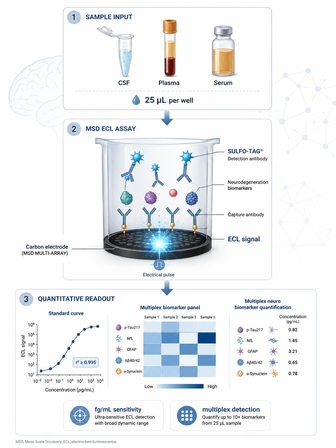
MSD's platform resolves this tension through spatially separated, electrically addressed detection spots within each well. Unlike fluorescence-based multiplexing, where overlapping emission spectra require compensation gating and sacrifice sensitivity at higher plex levels, each MSD analyte occupies a discrete electrode — addressed sequentially, with zero optical cross-talk. The result: ELISA-comparable sensitivity in a multiplex format, with 25 µL sample consumption regardless of plex level.
70+ pre-configured MSD panels for cytokine, chemokine, and immune mediator profiling — from focused human inflammation panels to broad biomarker discovery platforms. Available as full-service (sample-to-data) or kit-only.
| Panel / Service | Analytes | Species | Type | Sensitivity |
|---|---|---|---|---|
| MSD Human Inflammation 10-Plex | IFN-γ, IL-1β, IL-2, IL-4, IL-6, IL-8, IL-10, IL-12p70, IL-13, TNF-α | Human | Service / Kit | pg/mL |
| MSD Human Cytokine 30-Plex | GM-CSF, IFN-γ, IL-1α, IL-1β, IL-2, IL-4, IL-5, IL-6, IL-7, IL-8, IL-10, IL-12/IL-23p40, IL-12p70, IL-13, IL-15, IL-16, IL-17A, TNF-α, TNF-β, VEGF-A, Eotaxin, Eotaxin-3, IP-10, MCP-1, MCP-4, MDC, MIP-1α, MIP-1β, TARC | Human | Service / Kit | pg/mL |
| MSD Human Cytokine 36-Plex | GM-CSF, IFN-γ, IL-1α, IL-1β, IL-2, IL-4, IL-5, IL-6, IL-7, IL-8, IL-10, IL-12/IL-23p40, IL-12p70, IL-13, IL-15, IL-16, IL-17A, IL-21, IL-22, IL-23, IL-27, IL-31, TNF-α, TNF-β, VEGF-A, Eotaxin, Eotaxin-3, IP-10, MCP-1, MCP-4, MDC, MIP-1α, MIP-1β, MIP-3α, TARC | Human | Service / Kit | pg/mL |
| MSD Human Biomarker 54-Plex | CRP, Eotaxin, Eotaxin-3, FGF (basic), GM-CSF, ICAM-1, IFN-γ, IL-1α, IL-1β, IL-1RA, IL-2, IL-3, IL-4, IL-5, IL-6, IL-7, IL-8, IL-9, IL-10, IL-12/IL-23p40, IL-12p70, IL-13, IL-15, IL-16, IL-17A, IL-17A/F, IL-17B, IL-17C, IL-17D, IL-21, IL-22, IL-23, IL-27, IL-31, IP-10, MCP-1, MCP-4, MDC, MIP-1α, MIP-1β, MIP-3α, PlGF, SAA, TARC, Tie-2, TNF-α, TNF-β, TSLP, VCAM-1, VEGF-A, VEGF-C, VEGF-D, VEGFR-1/Flt-1 | Human | Service / Kit | pg/mL |
| MSD Human Chemokine 11-Plex (Panel 1) | Eotaxin, Eotaxin-3, IL-8, IP-10, MCP-1, MCP-4, MDC, MIP-1α, MIP-1β, TARC | Human | Service / Kit | pg/mL |
| MSD Human Chemokine 11-Plex (Panel 2) | CTACK, ENA-78, Fractalkine, GRO-α, I-309, I-TAC, MIF, MIP-3α, MIP-3β, MIP-5, SDF-1α | Human | Service / Kit | pg/mL |
| MSD Human Chemokine 13-Plex | Eotaxin, Eotaxin-2, Eotaxin-3, IL-8, IP-10, MCP-1, MCP-2, MCP-3, MCP-4, MDC, MIP-1α, MIP-1β, TARC | Human | Service / Kit | pg/mL |
| MSD Human Th1/Th2 10-Plex | IFN-γ, IL-1β, IL-2, IL-4, IL-5, IL-8, IL-10, IL-12p70, IL-13, TNF-α | Human | Service / Kit | pg/mL |
| MSD Human Th17 7/9/10-Plex | IL-17A, IL-17F, IL-17A/F, IL-21, IL-22, IL-23, IL-31, TNF-α, IL-6, GM-CSF | Human | Service / Kit | pg/mL |
| MSD Human T-Cell 14-Plex | GM-CSF, IFN-γ, IL-2, IL-4, IL-5, IL-6, IL-10, IL-12p70, IL-13, IL-17A, IL-21, IL-22, IL-23, TNF-α | Human | Service / Kit | pg/mL |
| MSD Human Inflammation 9-Plex (Ultrasensitive) | IFN-γ, IL-1β, IL-2, IL-4, IL-6, IL-10, IL-12p70, IL-17A, TNF-α | Human | Service / Kit | fg/mL (S-PLEX) |
| MSD Mouse Inflammation 10-Plex | IFN-γ, IL-1β, IL-2, IL-4, IL-5, IL-6, IL-10, IL-12p70, KC/GRO, TNF-α | Mouse | Service / Kit | pg/mL |
| MSD Mouse Cytokine 19-Plex | 19 analytes: IFN-γ, IL-1β, IL-2, IL-4, IL-5, IL-6, IL-9, IL-10, IL-12p70, IL-15, IL-17A/F, IL-27p28, IL-33, IP-10, KC/GRO, MCP-1, MIP-1α, MIP-2, TNF-α | Mouse | Service / Kit | pg/mL |
| MSD NHP Cytokine 24-Plex | Eotaxin-3, GM-CSF, IFN-γ, IL-1β, IL-2, IL-5, IL-6, IL-7, IL-8, IL-10, IL-12/IL-23p40, IL-15, IL-16, IL-17A, IP-10, MCP-1, MCP-4, MDC, MIP-1α, MIP-1β, TARC, TNF-β, VEGF-A | NHP | Service / Kit | pg/mL |
| MSD U-PLEX Custom Cytokine Panel | Select any 2–10 analytes from available targets; linker-coupled antibody format for full configuration flexibility | Multi-Species | Service | pg/mL–fg/mL |
Panel availability should be confirmed during project consultation. The panels listed represent core configurations in our MSD assay portfolio — additional plex levels, species variants, and custom combinations available upon request. Reagent kits and service-only options are both available. All services are for research use only.
Start with your research objective — we'll match the optimal panel configuration.
4–10 Plex V-PLEX
Core pro-inflammatory cytokine panels (IFN-γ, IL-1β, IL-6, TNF-α + key interleukins). Ideal for hypothesis-driven studies, treatment response monitoring, and routine immune characterization.
30–36 Plex V-PLEX
Comprehensive cytokine + chemokine coverage for systems-level immune characterization. Covers Th1, Th2, Th17, and chemokine axes simultaneously for discovery and deep phenotyping.
54-Plex V-PLEX
The flagship 54-plex panel: cytokines, chemokines, growth factors, vascular markers, acute-phase proteins, and metabolic markers — the broadest single-well multiplex for human biomarker discovery.
11–13 Plex V-PLEX
Dedicated chemokine panels for immune cell trafficking, migration, and chemotaxis research. Two complementary 11-plex panels or combined 13-plex for extended coverage.
S-PLEX fg/mL
fg/mL-level sensitivity for cytokines present at ultra-low concentrations — ideal for precious samples, early biomarker signals, and analytes below the detection floor of standard assays.
U-PLEX 2–10 Plex
Design your own panel from available targets. Linker-coupled antibodies provide full flexibility — no custom development timeline. Our scientific team advises on analyte compatibility.
Not sure which panel fits your study? Contact our scientific team for a free consultation — we'll help you design the optimal assay configuration for your research objectives.
Why electrochemiluminescence outperforms fluorescent bead-based platforms for simultaneous cytokine and chemokine quantification.
Cytokines span an extraordinary concentration range — from sub-pg/mL (IL-4, IL-5) to ng/mL (acute-phase cytokines) — and exhibit complex cross-reactivity patterns due to shared receptor subunits and structural homology within cytokine families (IL-1 family, IL-6 family, chemokine CC/CXC subfamilies). Fluorescent bead-based platforms address the multiplex need but introduce spectral overlap at moderate-to-high plex densities, requiring compensation algorithms that propagate uncertainty into low-abundance cytokine measurements — precisely where precision matters most.
A standardized process from study design consultation through data delivery — optimized for multiplex cytokine and chemokine panels.
Discuss your research objectives, matrix type (serum/plasma/CSF/supernatant), species, and analytes of interest. We help select the optimal V-PLEX panel or configure a custom U-PLEX combination, advise on sample volume requirements, and finalize the experimental design.
Ship your samples following our matrix-specific collection guidelines. Minimum 25 µL per sample regardless of plex level. Samples accessioned, aliquoted, and stored at −80°C with full chain-of-custody documentation.
Assays performed on MESO QuickPlex SQ 120 or MESO SECTOR S 600 instruments. Each plate includes multi-analyte 8-point standard curves, three-level QC controls in duplicate, and blank wells. All runs follow documented, standardized protocols.
Raw ECL signals acquired via MESO Discovery Workbench. QC acceptance criteria: intra-plate CV <10%, inter-plate CV <15%, standard curve R² ≥ 0.99 per analyte. Values outside calibration range flagged for re-analysis at appropriate dilution.
Comprehensive report: analyte concentrations for all targets, QC metrics per analyte, standard curve plots, raw ECL signal data, and a methods summary formatted for publication. Data delivered in Excel and PDF formats.
Matrix-specific collection guidance for cytokine and chemokine analysis.
Collect in EDTA tubes. Centrifuge within 30 min at 1,500×g for 10 min at 4°C. Transfer supernatant, aliquot into polypropylene cryovials, and freeze at −80°C. EDTA plasma is preferred for most cytokine panels — the clotting process in serum collection can introduce pre-analytical cytokine variability. Heparin and citrate also acceptable for most panels.
Collect in serum separator tubes (SST). Allow 30 min clotting at RT. Centrifuge at 1,500×g for 10 min at 4°C. Aliquot and freeze at −80°C. Consistent clotting time is critical — prolonged clotting releases platelet-derived cytokines (TGF-β1, PDGF-BB) that may bias measurements. Note matrix type in metadata; do not mix serum and plasma within a study.
Collect in polypropylene tubes. Centrifuge at 2,000×g for 10 min at 4°C. Aliquot immediately into polypropylene cryovials and store at −80°C. CSF is low-protein and low-volume — 25 µL per well is especially valuable given limited CSF availability. Supported by all MSD cytokine and chemokine panels with documented matrix performance data.
Centrifuge at 300×g for 5 min to remove cells, then at 2,000×g for 10 min to remove debris. Aliquot and freeze at −80°C. Note medium composition — serum-containing media introduces bovine cytokines that may cross-react with certain assays. Serum-free or low-serum conditions recommended where feasible. Include media-only controls for background subtraction.
Flash-freeze tissue at collection. Homogenize in appropriate lysis buffer with protease inhibitors. Centrifuge at 14,000×g for 10 min at 4°C to clear debris. Aliquot supernatant and freeze at −80°C. Total protein normalization recommended — report cytokine concentrations per mg total protein. Lysis buffer compatibility verified during project consultation.
Bronchoalveolar lavage (BAL), synovial fluid, urine, and other non-standard matrices are supported on consultation. Matrix-specific validation may be required — contact our scientific team to discuss feasibility for your specific matrix. Dilution linearity and spike recovery verification recommended for matrices not previously characterized with your panel of interest.
Shipping: All samples should be shipped on dry ice via overnight courier. We provide sample collection kits (tubes, labels, shipping containers) upon request. Avoid repeated freeze-thaw cycles — cytokines are susceptible to degradation with each cycle. Single-use aliquots strongly recommended. Record freeze-thaw count on each aliquot. Hemolyzed or lipemic samples may affect certain analytes and will be flagged during QC review rather than silently excluded. For multi-site studies, we can provide harmonized collection SOPs for distribution to all participating sites.
Every MSD cytokine assay engagement includes a complete data package designed for research documentation and publication.
Analyte concentrations for all targets with per-analyte QC metrics (intra-plate CV, inter-plate CV), standard curve parameters (R², back-fit accuracy), and LOD/LLOQ values. Delivered in Excel (.xlsx) and PDF formats.
Raw ECL signal values for every well, per-analyte standard curve plots with fitted curves, QC sample trend charts, and plate layout maps for full traceability. Compatible with downstream analysis in R, Python, or GraphPad Prism.
A detailed methods section describing assay protocol, reagents, instrument settings, and QC acceptance criteria — structured for direct use in manuscript Methods sections or supplementary materials.
Head-to-head technical comparison: the analytical factors that determine cytokine data quality in research applications.
Each analyte occupies a discrete electrode spot, electrically addressed in sequence. Fluorescent bead-based systems (Luminex) deconvolve overlapping emission spectra — a process that introduces compensation error and disproportionately affects low-abundance cytokines. MSD's spatial separation eliminates this problem entirely: 4-plex or 54-plex, every analyte gets an independent signal channel.
Measure IL-6 at ng/mL and IL-4 at sub-pg/mL in the same well under identical conditions. Fluorescent bead platforms typically offer 2–3 logs — requiring dilution adjustments that introduce pre-analytical variability when high- and low-abundance cytokines are measured together.
SULFO-TAG labels emit light only upon electrical stimulation at the electrode surface — no excitation light source means zero autofluorescence from serum, plasma, or tissue culture media. This is critical for low-pg/mL cytokine quantification, where matrix autofluorescence in optical systems can exceed the analyte signal.
MSD's electrical readout acquires all 96 wells in ~70 seconds. Fluorescent bead systems require 45–60 minutes of sequential laser interrogation per plate — a throughput difference that matters when running large cohort studies or multiple panels per sample.
A 54-plex consumes the same sample volume as a single ELISA well. Sample volume is independent of the number of analytes measured — preserving precious longitudinal aliquots, pediatric samples, and CSF for additional analyses.
The same ECL detection chemistry across V-PLEX, U-PLEX, and S-PLEX product lines means you can discover biomarkers with the 54-Plex, narrow to a focused U-PLEX panel, and validate with S-PLEX sensitivity — all on one instrument family, eliminating cross-platform variability.
V-PLEX panels are manufacturer-pre-validated multiplex kits with full analytical documentation and lot-to-lot bridging data — ideal for studies requiring documented, reproducible performance across timepoints. U-PLEX enables custom panel configuration: select any 2–10 analytes from available targets using linker-coupled antibodies, providing flexibility without custom development timelines. S-PLEX is the ultrasensitive single-analyte format achieving fg/mL-level detection for low-abundance cytokines through enhanced signal amplification. The optimal choice depends on your research needs: V-PLEX for validated, ready-to-use panels; U-PLEX for custom combinations; S-PLEX when standard sensitivity is insufficient.
MSD uses spatially separated electrode spots addressed electrically in sequence — there is no spectral overlap, no compensation gating, and no signal bleed-through between analytes regardless of plex level. Fluorescent bead-based platforms (Luminex xMAP) rely on spectral deconvolution of overlapping fluorophore emissions, which becomes increasingly complex at higher plex densities and can compromise low-abundance cytokine measurements. MSD also offers wider dynamic range (4–6 logs vs. 2–3 logs for Luminex), faster read time (~70 seconds vs. 45–60 minutes per plate), and background-free detection via electrical excitation — eliminating the autofluorescence interference common in serum and plasma. For applications requiring clean, precise cytokine quantification at moderate-to-high multiplex levels, MSD's spatial separation architecture provides distinct analytical advantages.
EDTA plasma is generally preferred for cytokine panels — it avoids the clotting-associated release of platelet-derived cytokines (TGF-β1, PDGF-BB, PF4) that can confound serum measurements. Serum is acceptable for most panels but may exhibit higher baseline concentrations of certain cytokines due to the clotting process. Cell culture supernatant should be centrifuged to remove cells and debris before freezing. The critical requirement is matrix consistency within a study — never mix serum and plasma in the same dataset. For cross-study comparisons, note the collection matrix in your metadata. Our scientific team can advise on matrix selection during study design based on your specific analytes of interest and published matrix performance data.
Yes. Many MSD panels already integrate cytokines, chemokines, growth factors, and soluble receptors in pre-configured formats — the Human Biomarker 54-Plex is the most comprehensive example, spanning 54 analytes across multiple functional categories. Through the U-PLEX custom platform, you can select any 2–10 targets from the available catalog to create a study-specific panel. Our scientific team can evaluate analyte compatibility, advise on expected concentration ranges, and help optimize the multiplex configuration for your research objectives. For targets not covered by existing V-PLEX or U-PLEX options, custom R-PLEX development is available.
Every MSD cytokine assay includes comprehensive QC documentation: (1) 8-point standard curve with R² and back-fit accuracy per analyte; (2) three-level QC samples (low, medium, high) run in duplicate on each plate with pre-defined acceptance windows; (3) intra-plate CV for each analyte; (4) inter-plate CV for multi-plate studies; (5) LOD and LLOQ determination per analyte; (6) raw ECL signal data for every well; (7) dilution linearity verification for the sample matrix; (8) spike recovery data (available for V-PLEX panels). All QC results are reported transparently — flagged values are annotated with rationale and retained in the dataset with full context.
Online Inquiry
×