
- Services Overview
- Analytes Details
- FAQ
Human cell proliferation and metastasis are two fundamental processes that play a critical role in both physiological and pathological contexts, particularly in cancer progression. While proliferation refers to the regulated process of cell growth and division, metastasis describes the spread of cancer cells from their original site to distant parts of the body, forming secondary tumors. Both of these processes are tightly controlled under normal conditions, but dysregulation can lead to tumorigenesis, aggressive malignancies, and metastasis — the hallmark of cancer lethality.
What is Human Cell Proliferation?
Cell proliferation is the process through which cells divide and multiply. This process is vital for tissue growth, repair, and maintenance. The process is regulated by several molecular pathways, including the cell cycle and various growth factors, to ensure normal function. Aberrations in cell proliferation mechanisms, such as unchecked growth signals or failure to trigger cell death (apoptosis), are hallmarks of cancerous cells. The unchecked division of these abnormal cells contributes to the development of tumors and ultimately cancer progression.
What is Metastasis?
Metastasis is the process by which cancer cells spread from the primary tumor to distant organs. This multi-step process involves cancer cells detaching from the primary tumor, invading surrounding tissues, entering the bloodstream (or lymphatic system), and eventually colonizing other parts of the body. Once metastatic cells establish themselves in a new environment, they begin to proliferate and form secondary tumors, often leading to poor patient prognosis.
Human Cell Proliferation and Metastasis Panel at Creative Proteomics
Creative Proteomics offers Human Cell Proliferation and Metastasis 12-plex/13plex Panel analysis services utilizing Luminex xMAP technology. Our panels allow for the simultaneous quantification of multiple biomarkers during cell growth, migration, and metastasis with minimal sample volume. This multiplexed approach provides a holistic view of cellular dynamics, aiding in the study of cancer progression, tumor aggressiveness and metastatic potential.
Detection Method
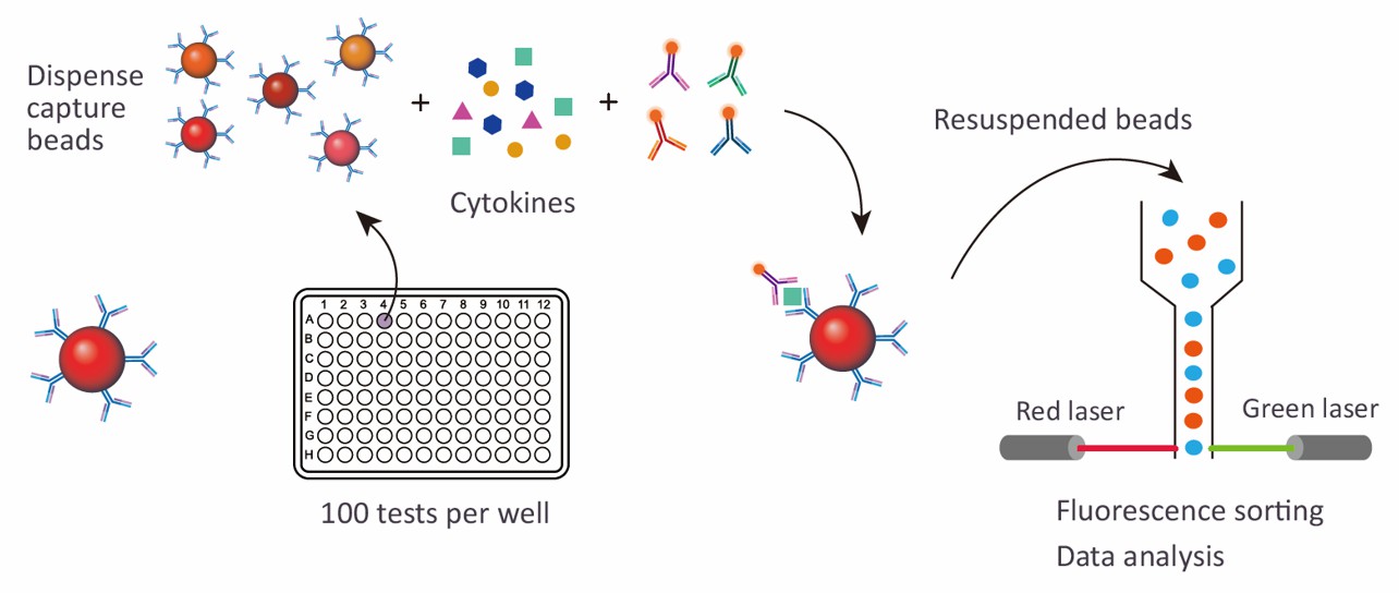
Magnetic bead-based Luminex multiplex assay
Species
Human
Analytes Detected
| Species | Specification | Protein Targets | Applications | Price |
|---|---|---|---|---|
| Human | Human Cell Proliferation and Metastasis 12-plex Panel | Beta-2-microglobulin (B2M), Cathepsin D, CEA (CEACAM-5), EGFR (ErbB1), Haptoglobin, HGFR (c-Met), IGFBP-2, IGFBP-3, MIA, MIP-4 (CCL18), Periostin (OSF-2), VE-cadherin | Ideal for studying cancer cell growth, proliferation, and metastatic spread | +Inquiry |
| Human | Human Cell Proliferation and Metastasis 13-plex Panel | AFP (Alpha-fetoprotein), ADAM12, Beta-catenin, CD171 (NCAM-L1), CD66f (PSBG-1), Galectin-1, hCG, HO-1 (Heme oxygenase 1), IGFBP-1, N-cadherin, Nectin-4, PAPP-A, Thrombomodulin (TM) 1 | Ideal for studying tumor biology, cell adhesion, and invasion mechanisms | +Inquiry |
Advantages of the Human Cell Proliferation and Metastasis Luminex Assay
- Multiplexing Capability: Unlike conventional methods such as ELISA or Western blot, which measure only a single analyte per assay, the Luminex xMAP platform enables the simultaneous detection of multiple biomarkers in a single sample. This provides a more comprehensive understanding of complex biological processes, such as cancer cell proliferation and metastasis, while saving valuable time, reagents, and sample volume.
- High Sensitivity and Specificity: The Luminex xMAP assay is designed for highly sensitive and specific detection, allowing the quantification of low-abundance biomarkers that are critical in early cancer development or metastasis. This sensitivity ensures the detection of subtle molecular changes, facilitating earlier diagnosis and a deeper understanding of disease progression.
- High-Throughput Efficiency: The Luminex technology supports high-throughput analysis, making it possible to analyze hundreds of samples and biomarkers simultaneously. This is particularly valuable for large-scale studies, clinical trials, and drug screening, where efficiency and data throughput are essential.
- Comprehensive Pathway Analysis: The ability to monitor multiple biomarkers simultaneously enables researchers to gain a holistic view of key pathways involved in cell proliferation and metastasis. The assay can detect a variety of proteins, including growth factors, adhesion molecules, and matrix metalloproteinases, providing comprehensive insights into tumor behavior and metastatic potential.
- Customizability: Creative Proteomics offers customizable panels, allowing researchers to tailor the assay to meet their specific needs. Whether focusing on a particular set of proliferation markers or investigating a wider range of metastasis-related proteins, the assay can be adapted to suit diverse research objectives.
- Cost and Time Efficiency: By allowing multiplex analysis from a single sample, the Luminex assay significantly reduces both cost and time compared to running multiple single-analyte assays. This makes it an ideal solution for projects requiring rapid data generation with limited resources.
- Minimal Sample Volume Requirement: The Luminex assay requires only minimal sample volumes, which is particularly advantageous when dealing with scarce or precious samples, such as clinical biopsies or specific tissue extracts.
Sample Requirements for Human Cell Proliferation and Metastasis Luminex Panel
| Sample Type | Volume Required | Handling Instructions | Storage Conditions |
|---|---|---|---|
| Serum | 50 - 100 µL | Collect in a serum separator tube, centrifuge within 30 minutes of collection | Store at -80°C |
| Plasma (EDTA or Heparin) | 50 - 100 µL | Collect in an anticoagulant tube, centrifuge within 30 minutes | Store at -80°C |
| Cell Supernatant | 50 - 100 µL | Collect and centrifuge to remove debris | Store at -80°C |
| Tissue Homogenate | 50 - 200 µL | Homogenize in appropriate lysis buffer, centrifuge | Store at -80°C |
| Urine | 100 - 200 µL | Centrifuge to remove particulates | Store at -80°C |
| Cell Lysate | 50 - 100 µL | Lyse cells using appropriate lysis buffer, centrifuge to clear debris | Store at -80°C |
| Bronchoalveolar Lavage (BAL) | 100 - 200 µL | Centrifuge to remove cells and debris, aliquot supernatant | Store at -80°C |
| Cerebrospinal Fluid (CSF) | 50 - 100 µL | Collect in sterile tubes, centrifuge within 30 minutes | Store at -80°C |
| Saliva | 100 - 200 µL | Collect using standard saliva collection protocol, centrifuge | Store at -80°C |
| Pleural Effusion | 100 - 200 µL | Centrifuge to remove cells and debris | Store at -80°C |
| Ascites Fluid | 100 - 200 µL | Centrifuge to remove cells and debris | Store at -80°C |
| Sample Type | Volume Required | Handling Instructions | Storage Conditions |
| Serum | 50 - 100 µL | Collect in a serum separator tube, centrifuge within 30 minutes of collection | Store at -80°C |
| Plasma (EDTA or Heparin) | 50 - 100 µL | Collect in an anticoagulant tube, centrifuge within 30 minutes | Store at -80°C |
| Cell Supernatant | 50 - 100 µL | Collect and centrifuge to remove debris | Store at -80°C |
| Tissue Homogenate | 50 - 200 µL | Homogenize in appropriate lysis buffer, centrifuge | Store at -80°C |
| Urine | 100 - 200 µL | Centrifuge to remove particulates | Store at -80°C |
Application of Human Cell Proliferation and Metastasis Panel
- Cancer Biology Research
Understanding Tumor Growth: The panel allows researchers to quantify biomarkers related to cell proliferation, helping elucidate the mechanisms driving tumor growth and progression.
Studying Metastatic Mechanisms: By measuring proteins involved in cell migration and invasion, the panel provides insights into the biological processes underlying metastasis.
- Biomarker Discovery and Validation
Identification of New Biomarkers: The multiplex approach enables the discovery of novel biomarkers associated with tumor behavior and patient outcomes.
Validation of Existing Biomarkers: Researchers can validate previously identified biomarkers in various cancer types, enhancing their potential for clinical use.
- Therapeutic Development
Target Identification: The panel can help identify potential therapeutic targets by assessing the expression of proteins involved in cell proliferation and metastasis.
Drug Efficacy Studies: It enables the evaluation of drug effects on specific biomarkers, assisting in the development and testing of new cancer therapies.
- Comparative Studies
Comparing Tumor Microenvironments: Researchers can use the panel to analyze differences in biomarker expression between primary tumors and metastases, providing insights into the tumor microenvironment and its role in cancer progression.
Evaluating Variations Across Cancer Types: The ability to assess multiple biomarkers allows for comparative studies across different types of cancer, facilitating the understanding of disease-specific mechanisms.
- Basic Research
Exploring Cell Signaling Pathways: The panel can be utilized to investigate key signaling pathways involved in cell proliferation and metastasis, providing valuable data for basic cancer research.
In addition to preconfigured panels, we also offer customized analysis services. You can customize your own panel through our customization tool, or directly email us the targets you are interested in. A professional will contact you to discuss the feasibility of customization. We look forward to working with you!
| Protein Target | Description |
|---|---|
| Beta-2-microglobulin (B2M) | A protein associated with immune response and tumor progression; elevated levels may indicate poor prognosis in various cancers. |
| Cathepsin D | A cysteine protease involved in cancer cell invasion and metastasis, it plays a role in the degradation of extracellular matrix components. |
| CEA (CEACAM-5) | Carcinoembryonic antigen, a glycoprotein associated with colorectal and other cancers; often used as a tumor marker in clinical settings. |
| EGFR (ErbB1) | Epidermal growth factor receptor, a key player in cell proliferation and survival; its overexpression is linked to various cancers, including lung cancer. |
| Haptoglobin | A plasma protein that binds free hemoglobin; it is involved in inflammation and is associated with poor outcomes in cancer patients. |
| HGFR (c-Met) | Hepatocyte growth factor receptor, involved in cell growth, motility, and invasion; overactivation is linked to metastatic behavior in cancers. |
| IGFBP-2 | Insulin-like growth factor-binding protein 2, which regulates IGF activity; altered levels are associated with cancer progression. |
| IGFBP-3 | Another member of the IGF-binding proteins, it has both growth-promoting and growth-inhibiting activities, influencing cancer cell dynamics. |
| MIA | Melanoma inhibitory activity, a protein linked to tumor progression; elevated levels can indicate aggressive tumor behavior in melanoma. |
| MIP-4 (CCL18) | A chemokine involved in the recruitment of immune cells to tumors; associated with a pro-tumor microenvironment. |
| Periostin (OSF-2) | An extracellular matrix protein involved in cell adhesion and migration; its expression is linked to tumor growth and metastasis. |
| VE-cadherin | A key component of endothelial cell junctions; its regulation is crucial for maintaining vascular integrity and is implicated in tumor angiogenesis. |
| AFP (Alpha-fetoprotein) | A glycoprotein typically elevated in liver cancer and germ cell tumors; it serves as a biomarker for diagnosis and monitoring treatment response. |
| ADAM12 | A disintegrin and metalloproteinase involved in cell adhesion and migration; it plays a role in the invasive properties of cancer cells. |
| Beta-catenin | A key player in the Wnt signaling pathway, involved in cell proliferation and differentiation; its dysregulation is often seen in cancers. |
| CD171 (NCAM-L1) | Neural cell adhesion molecule involved in cell adhesion and migration; its expression is linked to tumor invasiveness and metastasis. |
| CD66f (PSBG-1) | A glycoprotein associated with immune cell activation; elevated levels are often found in various cancer types, indicating tumor progression. |
| Galectin-1 | A carbohydrate-binding protein that modulates cell adhesion and immune response; it is associated with tumor progression and metastasis. |
| hCG | Human chorionic gonadotropin, typically elevated in germ cell tumors; it is used as a tumor marker in clinical settings. |
| HO-1 (Heme oxygenase 1) | An enzyme that provides cytoprotection against oxidative stress; its elevated expression is linked to cancer progression and resistance to therapy. |
| IGFBP-1 | Insulin-like growth factor-binding protein 1, which can modulate cell growth and survival; its levels may be altered in various cancers. |
| N-cadherin | A cadherin involved in cell adhesion; its expression is associated with epithelial-mesenchymal transition (EMT) and cancer metastasis. |
| Nectin-4 | A cell adhesion molecule involved in the regulation of cell-cell interactions; its expression can influence tumor aggressiveness. |
| PAPP-A | Pregnancy-associated plasma protein A, linked to tissue remodeling and angiogenesis; altered levels can be indicative of certain malignancies. |
| Thrombomodulin (TM) 1 | An integral membrane protein that acts as a cofactor for thrombin; its expression is associated with endothelial function and cancer progression. |
What is the sensitivity of the Luminex assay used in this panel?
The Luminex xMAP technology employed in the Human Cell Proliferation and Metastasis Panel offers high sensitivity, capable of detecting low-abundance biomarkers in the picogram per milliliter range. This allows researchers to identify subtle changes in biomarker levels that may be indicative of early tumor development or progression.
How are the results reported, and what statistical analyses are provided?
Results from the Human Cell Proliferation and Metastasis Panel are reported in a comprehensive format that includes quantitative measurements for each target biomarker. Creative Proteomics provides statistical analysis as needed, including mean values, standard deviations, and comparison with control samples. Custom analytical approaches, such as correlation analyses or multivariate statistics, can also be performed to aid in interpreting the biological significance of the results.
What is the turnaround time for results?
The typical turnaround time for the Human Cell Proliferation and Metastasis Panel results is approximately 2-4 weeks, depending on the complexity of the analysis and the sample type. Urgent requests can often be accommodated; please contact us directly to discuss your timeline needs and we will do our best to meet your requirements.