- Services Overview
- Analytes Details
- FAQ
What is Mouse Angiogenesis?
Angiogenesis is the biological process of forming new blood vessels from existing ones, essential for various physiological functions such as embryonic development and tissue repair. However, dysregulation of angiogenesis is implicated in numerous diseases, including cancer and cardiovascular disorders.
In healthy tissues, angiogenesis is regulated by a balance of pro-angiogenic and anti-angiogenic factors. For instance, vascular endothelial growth factor (VEGF) promotes new blood vessel formation in response to hypoxia or injury. In contrast, tumors often exploit these pathways, leading to excessive blood vessel growth that facilitates cancer progression.
Mouse models are vital for studying angiogenesis due to their genetic and physiological similarities to humans. Researchers can manipulate specific genes, providing insights into the mechanisms of angiogenesis and its role in disease. Observing these processes in vivo yields valuable data that in vitro studies may not fully capture.
To understand the angiogenic response in mouse models, comprehensive analysis of multiple angiogenic factors is crucial. The Mouse Angiogenesis Panel is designed to assess various key biomarkers involved in this process, allowing researchers to gain a holistic view of angiogenesis in their models. This facilitates the identification of dysregulated pathways and the development of targeted interventions.
Mouse Angiogenesis Panel at Creative Proteomics
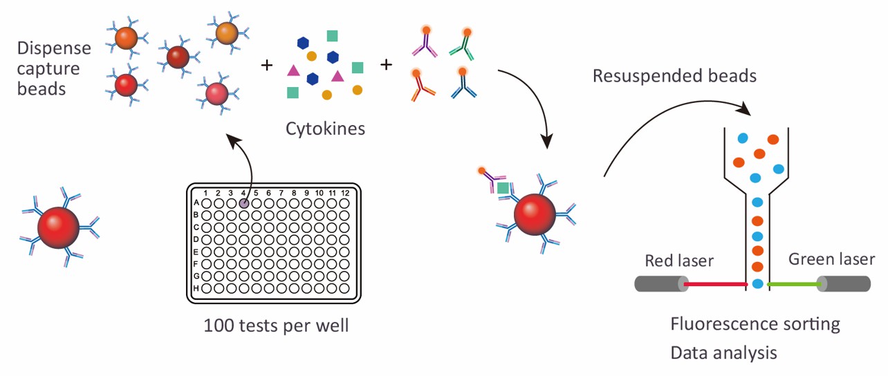
The Mouse Angiogenesis 8-plex/24-plex Panel service at Creative Proteomics offers multiplex assays to measure a range of angiogenic biomarkers. Utilizing advanced Luminex xMAP technology, these panels provide high-sensitivity and high-throughput analysis, enabling researchers to efficiently study angiogenesis in mouse models.
Detection Method
Magnetic bead-based Luminex multiplex assay
Species
Mouse
Analytes Detected
| Species | Specification | Protein Targets | Applications | Price |
|---|---|---|---|---|
| Mouse | Mouse Angiogenesis 8-plex Panel | HGF, IGFBP-1, IGFBP-3, IL-6R alpha, MMP-9, Osteopontin/OPN, VEGF, VEGFR2/KDR/Flk-1 | Ideal for analyzing angiogenic responses and vascular biology | +Inquiry |
| Mouse | Mouse Angiogenesis 24-plex Panel | Adiponectin/Acrp30, Endoglin/CD105, Angiopoietin-2, CCL2/JE/MCP-1, CCL3/MIP-1 alpha, CXCL10/IP-10/CRG-2, CXCL16, DPPIV/CD26, EGF, FGF basic/FGF2, G-CSF, GM-CSF, IL-1 alpha/IL-1F1, IL-1 beta/IL-1F2, IL-10, Leptin/OB, MMP-3, MMP-8, PDGF-AA, PDGF-BB, Prolactin, Serpin E1/PAI-1, TIMP-1, uPAR | Comprehensive profiling of angiogenesis and inflammatory factors | +Inquiry |
Advantages of the Mouse Angiogenesis Luminex Assay
- Multiplexing Capability: The assay can simultaneously quantify up to 100 analytes in a single sample, offering a comprehensive overview of angiogenic factors. This dramatically reduces the need for multiple separate assays, saving time and effort.
- High Sensitivity: Luminex assays boast a detection sensitivity down to pg/mL levels, ensuring accurate measurements of low-abundance proteins that are critical in early-stage angiogenesis or subtle biological changes.
- Reduced Sample Volume: Only 25–50 µL of sample is required per well, significantly less than traditional ELISAs, which often require 100 µL or more. This is crucial when working with limited or precious samples, such as tissue homogenates or cell culture supernatants.
- Cost Efficiency: Multiplexing reduces the cost per analyte tested by as much as 30% compared to running individual single-plex assays. This results in lower overall assay costs, while maintaining the integrity of high-throughput research.
- Throughput Capacity: The Luminex platform can process 96 samples per plate, making it ideal for large-scale studies. High throughput is essential for projects requiring multiple time points, treatments, or experimental conditions.

Sample Requirements for Mouse Angiogenesis Luminex Panel
| Sample Type | Volume Required | Storage Conditions | Notes |
|---|---|---|---|
| Serum | 25–50 µL | -80°C (long-term), -20°C (short-term) | Centrifuge at 2,000 x g for 10 min to remove debris before storage. |
| Plasma (EDTA, Heparin, Citrate) | 25–50 µL | -80°C (long-term), -20°C (short-term) | Centrifuge at 2,000 x g for 10 min to remove cells and platelets. |
| Cell Culture Supernatant | 100–200 µL | -80°C (long-term), -20°C (short-term) | Collect supernatant after 48–72 hours of incubation, remove debris by centrifugation. |
| Tissue Homogenates | 100–200 µg of protein | -80°C | Homogenize in RIPA buffer, centrifuge at 10,000 x g for 10 min, collect supernatant. |
| Urine | 100–200 µL | -80°C | Centrifuge at 1,000 x g for 10 min to remove particulates before freezing. |
| Bronchoalveolar Lavage Fluid (BALF) | 50–100 µL | -80°C | Centrifuge at 300 x g for 10 min, collect supernatant. |
| Lysates (Cell or Tissue) | 100–200 µg of protein | -80°C | Prepare lysates using appropriate lysis buffer, centrifuge to remove debris. |
| Synovial Fluid | 50–100 µL | -80°C | Centrifuge at 2,000 x g for 10 min to remove cells before freezing. |
Application of Mouse Angiogenesis Panel
- Oncology research
Angiogenesis is critical for tumor growth and metastasis. This panel quantifies key pro-angiogenic factors such as vascular endothelial growth factor, growth hormone and MMPs, helping researchers understand how tumors induce blood vessel formation. This in turn can help develop anti-angiogenesis targeted therapies that inhibit tumor angiogenesis and slow cancer progression.
- Cardiovascular and ischemic diseases
In ischemic heart disease and stroke, angiogenesis is associated with restoring blood flow and promoting tissue repair. This panel enables detailed analysis of angiogenic markers such as VEGFR2, PDGF and FGF, which can help develop therapies aimed at enhancing angiogenesis in damaged tissues, thereby improving recovery.
- Wound healing and tissue regeneration
Effective wound healing depends on the timely formation of new blood vessels to deliver nutrients and remove waste products. Mouse angiogenic panels help analyze the role of cytokines and growth factors such as angiopoietin-2 and TIMP-1, which are involved in the complex interactions that govern tissue regeneration.
- Metabolic disorders and obesity
Angiogenesis is associated with adipose tissue expansion and metabolic regulation. By analyzing markers such as leptin, adiponectin, and MCP-1, researchers can better understand the links between angiogenesis, inflammation, and metabolic disorders such as obesity and type 2 diabetes.
- Inflammation and autoimmune diseases
Angiogenesis is closely linked to inflammation in diseases such as rheumatoid arthritis and psoriasis. This panel provides detailed analysis of inflammatory and angiogenic cytokines, including IL-1 beta, IL-10 and VEGF, providing insight into how vascular abnormalities contribute to chronic inflammatory diseases.
In addition to preconfigured panels, we also offer customized analysis services. You can customize your own panel through our customization tool, or directly email us the targets you are interested in. A professional will contact you to discuss the feasibility of customization. We look forward to working with you!
| Protein Target | Description |
|---|---|
| HGF (Hepatocyte Growth Factor) | A growth factor involved in tissue regeneration and repair; promotes cell proliferation, angiogenesis, and wound healing. |
| IGFBP-1 (Insulin-like Growth Factor Binding Protein 1) | Regulates insulin-like growth factors, impacting cell growth, metabolism, and tissue repair processes. |
| IGFBP-3 (Insulin-like Growth Factor Binding Protein 3) | Modulates the availability and activity of insulin-like growth factors, playing a key role in cellular growth and metabolic regulation. |
| IL-6R alpha (Interleukin-6 Receptor alpha) | A receptor component crucial for IL-6 signaling, involved in immune responses, inflammation, and metabolic regulation. |
| MMP-9 (Matrix Metalloproteinase-9) | An enzyme involved in the breakdown of extracellular matrix, critical for tissue remodeling and angiogenesis. |
| Osteopontin/OPN | A multifunctional protein that regulates inflammation, immune responses, and extracellular matrix remodeling, often linked to chronic inflammatory diseases. |
| VEGF (Vascular Endothelial Growth Factor) | A critical angiogenic factor promoting new blood vessel formation, particularly important in cancer, wound healing, and vascular diseases. |
| VEGFR2/KDR/Flk-1 | A receptor for VEGF that mediates angiogenic signaling, playing a central role in endothelial cell proliferation and migration. |
| Adiponectin/Acrp30 | An adipokine with anti-inflammatory properties, crucial for regulating glucose levels and fatty acid breakdown. |
| Endoglin/CD105 | A glycoprotein involved in endothelial cell function and angiogenesis, particularly in vascular development and repair. |
| Angiopoietin-2 | A key regulator of blood vessel maturation and stability, playing a role in both promoting and inhibiting angiogenesis depending on the context. |
| CCL2/JE/MCP-1 | A chemokine that attracts monocytes to sites of inflammation, often elevated in obesity and chronic inflammatory conditions. |
| CCL3/MIP-1 alpha | A chemokine involved in the recruitment of immune cells to sites of infection and inflammation, contributing to immune responses. |
| CXCL10/IP-10/CRG-2 | A chemokine that recruits immune cells, particularly in response to inflammation or viral infections, also involved in tumor progression. |
| CXCL16 | A chemokine associated with inflammation and immune responses, involved in cardiovascular diseases and atherosclerosis. |
| DPPIV/CD26 | An enzyme that regulates glucose metabolism and immune responses, playing a role in metabolic disorders and immune regulation. |
| EGF (Epidermal Growth Factor) | A growth factor that stimulates cell growth, proliferation, and differentiation, playing an important role in wound healing and tissue regeneration. |
| FGF basic/FGF2 | A growth factor involved in angiogenesis, tissue regeneration, and wound healing, promoting the proliferation of endothelial cells. |
| G-CSF (Granulocyte Colony-Stimulating Factor) | Stimulates the production of neutrophils and promotes their mobilization, critical for immune response and tissue repair. |
| GM-CSF (Granulocyte-Macrophage Colony-Stimulating Factor) | A growth factor that stimulates the production of granulocytes and macrophages, important for immune defense and tissue regeneration. |
| IL-1 alpha/IL-1F1 | A pro-inflammatory cytokine involved in immune responses and inflammatory diseases, particularly in the acute phase of inflammation. |
| IL-1 beta/IL-1F2 | A key pro-inflammatory mediator involved in host defense, inflammation, and fever responses, contributing to chronic inflammatory conditions. |
| IL-10 | An anti-inflammatory cytokine that regulates immune responses, limiting excessive inflammation and autoimmunity. |
| Leptin/OB | A hormone produced by adipose tissue that regulates energy balance, appetite, and metabolism, closely linked to body fat levels and obesity. |
| MMP-3 | A matrix metalloproteinase involved in tissue remodeling, wound healing, and angiogenesis, critical for extracellular matrix breakdown. |
| MMP-8 | A collagenase that breaks down collagen, playing a role in tissue remodeling, wound healing, and inflammation. |
| PDGF-AA | A growth factor that stimulates cell growth, angiogenesis, and tissue repair, particularly important in wound healing and fibrosis. |
| PDGF-BB | A key factor in blood vessel formation and tissue repair, promoting the growth of endothelial cells and smooth muscle cells. |
| Prolactin | A hormone involved in lactation, reproduction, and immune regulation, with emerging roles in angiogenesis and tissue regeneration. |
| Serpin E1/PAI-1 | A protein that regulates fibrinolysis, playing a role in thrombosis and tissue repair, with elevated levels linked to obesity and metabolic disorders. |
| TIMP-1 (Tissue Inhibitor of Metalloproteinases-1) | An inhibitor of matrix metalloproteinases, involved in tissue remodeling and inflammation, often elevated in conditions such as fibrosis and cancer. |
| uPAR (Urokinase Plasminogen Activator Receptor) | A receptor that regulates proteolysis and cell migration, playing a key role in angiogenesis, tissue repair, and cancer metastasis. |
How sensitive is the Mouse Angiogenesis Panel? Can it detect low-abundance proteins?
The Luminex xMAP platform used in the panel provides high sensitivity, with detection levels down to the picogram per milliliter (pg/mL) range. This level of sensitivity allows for accurate quantification of low-abundance angiogenic factors that may play critical roles in early disease progression or subtle biological changes in mouse models.
What kind of controls should I include with my samples?
We recommend including both positive and negative controls in your assay. Positive controls may include samples known to have elevated levels of angiogenic markers, such as tumor or ischemic tissues. Negative controls could be from healthy, untreated samples. Internal reference standards are also advisable to normalize data across experimental conditions.

