- Services Overview
- Analytes Details
- FAQ
What is Mouse Oncology?
Mouse oncology refers to the use of mouse models in cancer research. Mice are commonly employed in this field because of their genetic, biological, and behavioral similarities to humans, making them ideal for studying tumor development, progression, and treatment responses.
In mouse oncology, researchers often utilize various types of models, including syngeneic models (where tumor cells from one mouse strain are implanted into genetically identical mice), xenograft models (where human tumor cells are implanted into immunocompromised mice), and transgenic models (genetically modified mice that express specific oncogenes or lack tumor suppressor genes).
The applications of mouse oncology are extensive. Researchers can investigate the molecular mechanisms underlying cancer, test the efficacy and safety of new therapies, and discover potential biomarkers for diagnosis and treatment. Despite some limitations—such as species differences that may affect translational relevance—mouse models remain a critical tool in cancer research, helping to advance our understanding of the disease and improve treatment options for patients.
Mouse Oncology Panels are specifically designed to profile and quantify various biological markers related to cancer in these models. These panels enable researchers to measure a wide range of cytokines, chemokines, and other analytes implicated in the development and progression of tumors.
Mouse Oncology Panel at Creative Proteomics
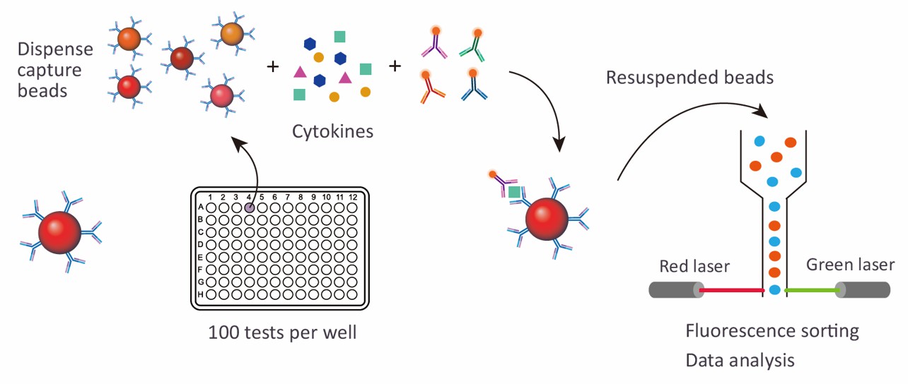
Creative Proteomics employs the Luminex xMAP technology to deliver high-throughput, multiplexed assays that ensure data integrity and consistency. This advanced platform allows simultaneous analysis of multiple proteins within a single sample, significantly enhancing research efficiency and discovering insights into cancer biology.
Detection Method
Magnetic bead-based Luminex multiplex assay
Species
Mouse
Analytes Detected
| Species | Specification | Protein Targets | Applications | Price |
|---|---|---|---|---|
| Mouse | Mouse Oncology 14-plex Panel | CCL8/MCP-2, G-CSF, HGF, IGFBP-1, IGFBP-3, IL-6, IL-6R alpha, Leptin/OB, MMP-9, Osteopontin/OPN, Periostin/OSF-2, TIMP-1, TNF-alpha, VEGFR2/KDR/Flk-1 | Suitable for assessing various cancer-related processes, including inflammation, tumor growth, and therapeutic efficacy. | +Inquiry |
| Mouse | Mouse Oncology 26-plex Panel | CCL3/MIP-1 alpha, ErbB2/Her2, G-CSF, HGF, IGFBP-3, IL-2, IL-6, IL-17E/IL-25, Kallikrein 3/PSA, Lumican, MIF, MMP-1, MMP-2, MMP-10, MMP-13, MSP/MST1, Myeloperoxidase/MPO, Osteoprotegerin/TNFRSF11B, Prolactin, TGF-alpha, Thymidine Kinase 1, Tie-2, TNF-alpha, TRAIL/TNFSF10, VCAM-1/CD106, VEGF | Ideal for comprehensive profiling of cancer biomarkers, including growth factors and immune modulators. | +Inquiry |
| Mouse | Mouse Oncology 27-plex Panel | Angiopoietin-2, BAFF/BLyS/TNFSF13B, CCL2/JE/MCP-1, CCL3/MIP-1 alpha, CCL5/RANTES, CCL7/MCP-3/MARC, CCL20/MIP-3 alpha, Chitinase 3-like 1, Dkk-1, EGF, Endoglin/CD105, Fas Ligand/TNFSF6, FGF basic/FGF2, GDF-15, GM-CSF, ICAM-1/CD54, IL-2, IL-17E/IL-25, M-CSF, MMP-2, MMP-3, MMP-8, PDGF-AA, Prolactin, Serpin E1/PAI-1, TWEAK/TNFSF12, VEGF | Enables detailed investigation of tumor microenvironments and the immune response to cancer therapies. | +Inquiry |
Advantages of the Mouse Oncology Luminex Assay
High Multiplexing Capability
- Broad Panel Selections: The assay can simultaneously quantify multiple analytes within a single sample. Panel options include the Mouse Oncology 14-plex, 26-plex, and 27-plex, catering to a range of research needs from targeted analysis to comprehensive profiling.
- Efficiency: This multiplexing reduces the time, cost, and sample volume required when compared to running multiple individual assays, thereby enhancing overall experimental efficiency.
Sensitivity and Specificity
- High Sensitivity: Luminex xMAP technology is known for its high sensitivity, allowing for the detection of low-abundance cytokines and growth factors, which are critical in cancer research.
- Specificity: The use of color-coded beads ensures precise identification and quantification of each target, minimizing cross-reactivity and ensuring reliable results.
Versatility and Application
- Wide Range of Applications: Suitable for various applications including the evaluation of therapeutic efficacy, studying tumor microenvironments, and understanding immune responses in cancer.
- Customizable: The assay can be tailored to include specific biomarkers relevant to your study, allowing researchers to focus on particular aspects of oncology.
Streamlined Workflow
- Automation-Ready: Compatible with automated systems, the Luminex Assay optimizes throughput while minimizing human error, making it ideal for high-throughput screening.
- Ease of Use: The user-friendly nature of the Luminex platform simplifies the assay setup and data analysis process, even for complex multi-analyte profiles.
Data Quality and Consistency
- Quantitative Precision: Provides quantitative results which are crucial for drawing meaningful conclusions from complex biological systems.
- Reproducibility: Assures high reproducibility across experiments, essential for longitudinal studies or collaborative research projects.

Sample Requirements for Mouse Oncology Luminex Panel
| Sample Type | Volume Required | Collection Notes |
|---|---|---|
| Serum | 50-100 µL | Collect in serum separator tubes, centrifuge, and store at -80°C |
| Plasma (EDTA/heparin) | 50-100 µL | Collect in anticoagulant tubes, centrifuge, and store at -80°C |
| Bone Tissue Homogenate | 100-200 mg | Homogenize in buffer, centrifuge to remove debris, and store at -80°C |
| Cell Culture Supernatant | 200 µL | Collect from confluent cultures, centrifuge, and store at -80°C |
| Urine | 500 µL | Collect fresh, centrifuge to remove particulates, and store at -80°C |
| Synovial Fluid | 100 µL | Collect aseptically, centrifuge if necessary, and store at -80°C |
| Bone Marrow Aspirate | 100 µL | Collect aseptically, centrifuge to separate cells if needed, and store at -80°C |
| Whole Blood | 100 µL | Collect with anticoagulant, gently mix, and store at -80°C for cellular analysis |
Application of Mouse Oncology Panel
- Cancer Biomarker Discovery
Translational Research: Bridging the gap between bench research and clinical application by translating findings from mouse models to human cancer studies.
Personalized Medicine: Supporting personalized treatment strategies by identifying specific protein targets that are modulated in individual cancer cases.
- Therapeutic Development
Assessing efficacy and safety of new oncology drugs by evaluating their impact on cancer-related pathways and biomarkers.
- Tumor Microenvironment Studies
Understanding the complex interactions within the tumor microenvironment, including immune cell infiltration and cytokine release.
- Translational Research
Bridging the gap between bench research and clinical application by translating findings from mouse models to human cancer studies.
- Personalized Medicine
Supporting personalized treatment strategies by identifying specific protein targets that are modulated in individual cancer cases.
In addition to preconfigured panels, we also offer customized analysis services. You can customize your own panel through our customization tool, or directly email us the targets you are interested in. A professional will contact you to discuss the feasibility of customization. We look forward to working with you!
Mouse Oncology 14-Plex Panel Protein Targets
| Protein Target | Description |
|---|---|
| CCL8 (MCP-2) | A chemokine involved in recruiting monocytes and basophils, playing a significant role in inflammation and tumor microenvironment modulation. |
| G-CSF | Granulocyte colony-stimulating factor, a cytokine that stimulates the production and release of neutrophils, contributing to immune responses against tumors. |
| HGF | Hepatocyte growth factor, involved in cell proliferation, migration, and angiogenesis, and linked to tumor growth and metastasis. |
| IGFBP-1 | Insulin-like growth factor-binding protein 1, which modulates the effects of insulin-like growth factors, impacting tumor cell proliferation and survival. |
| IGFBP-3 | A major carrier of insulin-like growth factors, influencing cell growth and apoptosis in various cancer types. |
| IL-6 | An inflammatory cytokine that promotes tumor growth, angiogenesis, and immune evasion in the tumor microenvironment. |
| IL-6R alpha | The alpha chain of the interleukin-6 receptor, facilitating IL-6 signaling pathways critical for cancer progression. |
| Leptin (OB) | A hormone involved in energy regulation, which also promotes cell proliferation and angiogenesis in tumor biology. |
| MMP-9 | Matrix metalloproteinase-9, an enzyme that degrades extracellular matrix components, facilitating tumor invasion and metastasis. |
| Osteopontin (OPN) | A glycoprotein involved in cell adhesion and migration, associated with tumor progression and metastasis. |
| Periostin (OSF-2) | An extracellular matrix protein involved in cell adhesion, migration, and angiogenesis, often upregulated in tumors. |
| TIMP-1 | Tissue inhibitor of metalloproteinases-1, which regulates MMP activity and is involved in tissue remodeling and tumor progression. |
| TNF-alpha | Tumor necrosis factor-alpha, a pro-inflammatory cytokine that plays a dual role in promoting and inhibiting tumor growth. |
| VEGFR2 (KDR/Flk-1) | A receptor for vascular endothelial growth factor, crucial for angiogenesis and the development of tumor vasculature. |
Mouse Oncology 26-Plex Panel Protein Targets
| Protein Target | Description |
|---|---|
| CCL3 (MIP-1 alpha) | A chemokine that recruits immune cells to the tumor site, influencing the inflammatory response and tumor microenvironment. |
| ErbB2 (Her2) | An oncogene associated with aggressive tumor behavior and poor prognosis, particularly in breast cancer. |
| G-CSF | Granulocyte colony-stimulating factor, facilitating the proliferation and differentiation of neutrophils in response to inflammation and tumors. |
| HGF | Hepatocyte growth factor, promoting cellular proliferation, migration, and angiogenesis, often implicated in cancer progression. |
| IGFBP-3 | Insulin-like growth factor-binding protein 3, modulating the effects of insulin-like growth factors on cell proliferation and apoptosis. |
| IL-2 | A cytokine critical for T cell proliferation and activation, playing a vital role in immune responses against tumors. |
| IL-6 | An inflammatory cytokine linked to tumor growth, angiogenesis, and immune evasion mechanisms. |
| IL-17E (IL-25) | A cytokine that can modulate immune responses and is associated with inflammation and tumor progression. |
| Kallikrein 3 (PSA) | Prostate-specific antigen, a serine protease involved in prostate cancer diagnosis and progression. |
| Lumican | An extracellular matrix protein that regulates cell proliferation and migration, influencing tumor growth and metastasis. |
| MIF | Macrophage migration inhibitory factor, a cytokine that promotes inflammation and tumor growth by inhibiting apoptosis. |
| MMP-1, MMP-2, MMP-10, MMP-13 | Matrix metalloproteinases involved in extracellular matrix remodeling, contributing to tumor invasion and metastasis. |
| MSP (MST1) | A protein involved in macrophage survival and differentiation, influencing tumor-associated inflammation. |
| Myeloperoxidase (MPO) | An enzyme produced by neutrophils, involved in the inflammatory response and associated with cancer progression. |
| Osteoprotegerin (TNFRSF11B) | A cytokine that inhibits osteoclastogenesis and is involved in bone metabolism, with implications in tumor-induced bone disease. |
| Prolactin | A hormone that can influence immune responses and promote tumor growth in certain cancer types. |
| TGF-alpha | A growth factor involved in epithelial cell proliferation and differentiation, often implicated in tumorigenesis. |
| Thymidine Kinase 1 | An enzyme associated with DNA synthesis and cell proliferation, often elevated in various malignancies. |
| Tie-2 | A receptor involved in angiogenesis and endothelial cell survival, playing a role in the development of the tumor vasculature. |
| TNF-alpha | A pro-inflammatory cytokine involved in systemic inflammation and capable of promoting or inhibiting tumor growth. |
| TRAIL (TNFSF10) | A cytokine that induces apoptosis in tumor cells while sparing normal cells, making it a target for cancer therapy. |
| VCAM-1 (CD106) | A cell adhesion molecule involved in leukocyte trafficking and inflammation, contributing to tumor progression. |
| VEGF | Vascular endothelial growth factor, a key regulator of angiogenesis that promotes the formation of new blood vessels in tumors. |
Mouse Oncology 27-Plex Panel Protein Targets
| Protein Target | Description |
|---|---|
| Angiopoietin-2 | A factor involved in angiogenesis, influencing vascular remodeling and tumor blood supply. |
| BAFF (BLyS, TNFSF13B) | A cytokine that regulates B cell development and survival, influencing immune responses in the tumor microenvironment. |
| CCL2 (JE, MCP-1) | A chemokine that recruits monocytes and other immune cells to the tumor site, impacting inflammation and tumor progression. |
| CCL3 (MIP-1 alpha) | A chemokine that promotes the recruitment of immune cells, influencing the tumor microenvironment. |
| CCL5 (RANTES) | A chemokine that plays a role in the recruitment of immune cells to inflamed tissues and tumors. |
| CCL7 (MCP-3, MARC) | A chemokine involved in the recruitment of monocytes and T cells, contributing to the immune response in tumors. |
| CCL20 (MIP-3 alpha) | A chemokine that attracts lymphocytes and plays a role in the immune response and tumor progression. |
| Chitinase 3-like 1 | A protein associated with inflammation and tissue remodeling, often upregulated in various cancers. |
| Dkk-1 | A Wnt signaling antagonist involved in the regulation of tumor growth and progression. |
| EGF | Epidermal growth factor, which stimulates cell growth, proliferation, and differentiation, often involved in cancer signaling pathways. |
| Endoglin (CD105) | A co-receptor for transforming growth factor-beta, involved in angiogenesis and implicated in tumor vasculature. |
| Fas Ligand (TNFSF6) | A protein that induces apoptosis in target cells, influencing immune evasion in tumors. |
| FGF basic (FGF2) | A growth factor involved in angiogenesis and tumor growth, playing a role in the tumor microenvironment. |
| GDF-15 | Growth differentiation factor 15, a cytokine associated with inflammation and cancer progression. |
| GM-CSF | Granulocyte-macrophage colony-stimulating factor, a cytokine that promotes the growth and differentiation of myeloid cells, influencing immune responses. |
| ICAM-1 (CD54) | An adhesion molecule involved in immune cell trafficking and activation, contributing to the inflammatory response in tumors. |
| IL-2 | A cytokine crucial for T cell proliferation and activation, playing a vital role in anti-tumor immunity. |
| IL-17E (IL-25) | A cytokine associated with immune modulation and inflammation, which can impact tumor progression. |
| M-CSF | Macrophage colony-stimulating factor, involved in the proliferation and differentiation of monocytes and macrophages, influencing tumor-associated inflammation. |
| MMP-2, MMP-3, MMP-8 | Matrix metalloproteinases that play roles in extracellular matrix remodeling and tumor invasion. |
| PDGF-AA | Platelet-derived growth factor, involved in cell proliferation and angiogenesis, and associated with tumor development. |
| Prolactin | A hormone that influences immune responses and can promote tumor growth in certain contexts. |
| Serpin E1 (PAI-1) | Plasminogen activator inhibitor-1, which regulates fibrinolysis and is involved in tissue remodeling and tumor progression. |
| TWEAK (TNFSF12) | A cytokine that can promote inflammation and influence tumor growth and immune responses. |
| VEGF | Vascular endothelial growth factor, a key regulator of angiogenesis that promotes the formation of new blood vessels in tumors. |
How does the Luminex xMAP technology work in the context of the mouse oncology panel?
The Luminex xMAP technology utilized in the mouse oncology panel employs color-coded beads coated with specific capture antibodies that are linked to unique fluorescent dyes. When a sample is introduced, the target proteins bind to the corresponding antibodies on the beads. After a washing step to remove unbound substances, a detection antibody conjugated with a fluorescent reporter is added. This forms a sandwich complex of capture antibody, target protein, and detection antibody. The bead's unique color identifies the target analyte, while the fluorescent signal quantifies its concentration. This multiplexing capability allows for the simultaneous measurement of multiple biomarkers, making it an efficient and high-throughput approach for profiling cancer-related processes.
How do you ensure the quality and reproducibility of results obtained from the mouse oncology panel?
Quality assurance and reproducibility of results from the Mouse Oncology Panel are prioritized through multiple measures. First, we adhere to strict standard operating procedures (SOPs) for sample handling, storage, and processing, ensuring consistency across experiments. Second, each assay undergoes rigorous validation using known standards and controls to establish a reliable range of detection and quantification. Third, we implement regular maintenance and calibration of the Luminex instruments to ensure accuracy and precision in measurements. Additionally, we utilize replicate samples and internal controls within each assay to monitor variability and provide confidence in the reproducibility of the data. By integrating these practices, we maintain high standards of quality that are critical for translating experimental findings into meaningful biological insights.
How do I interpret the results from the mouse oncology panel, and what statistical methods are recommended?
Interpretation of results from the Mouse Oncology Panel involves several steps, starting with data normalization to account for variations between samples. After processing the data, statistical methods such as ANOVA, t-tests, or regression analysis can be employed depending on the experimental design and objectives. For longitudinal studies, mixed-effects models may be suitable to account for repeated measures. It is essential to consider the biological context of the biomarkers being analyzed, as some may have overlapping functions or may be regulated by similar pathways. We recommend collaborating with a biostatistician or utilizing our consulting services to ensure robust data interpretation, especially when exploring complex interactions within the tumor microenvironment.

