- Services Overview
- Analytes Details
- FAQ
What is the Akt Pathway?
The Akt pathway, also known as the protein kinase B (PKB) pathway, plays a pivotal role in regulating cell survival, growth, and metabolism. Central to numerous cellular functions, Akt is activated via phosphatidylinositol 3-kinase (PI3K) in response to extracellular stimuli such as growth factors, cytokines, and insulin. Upon activation, Akt mediates downstream effects that promote cell proliferation, inhibit apoptosis, and contribute to cellular metabolism.
The dysregulation of the Akt pathway has been implicated in various diseases, including:
- Cancer: where aberrant Akt signaling promotes uncontrolled cell proliferation.
- Metabolic disorders: like Type 2 diabetes, where insulin signaling via Akt is disrupted.
- Autoimmune and inflammatory diseases: due to Akt's role in immune cell activation and survival.
Understanding and analyzing the components of the Akt pathway is essential for disease research, drug development, and personalized medicine approaches.
Why Analyze the Human Akt Pathway?
Disease Mechanism Elucidation: Analyzing the Akt pathway helps researchers understand disease progression mechanisms at the molecular level, particularly in cancer and inflammatory diseases.
Biomarker Discovery: Quantifying cytokine and protein expression involved in the Akt pathway can lead to the identification of diagnostic and prognostic biomarkers for various diseases.
Therapeutic Targeting: Given the Akt pathway's role in promoting cell survival and proliferation, it is an attractive target for drug development. Measuring pathway activation can be crucial for evaluating the efficacy of targeted therapies.
Immunological Research: The Akt pathway also plays a vital role in regulating immune responses, making it valuable for research into immune modulation and inflammation.
Human Akt Pathway Panel at Creative Proteomics
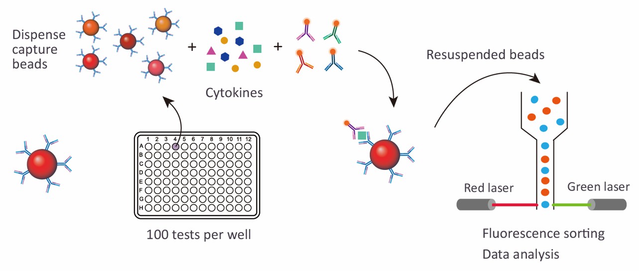
At Creative Proteomics, we utilize advanced Luminex xMAP technology to offer our specialized Human Akt Pathway 8-plex Panel. This panel is designed to provide a high-throughput, multiplexed analysis of key proteins involved in the Akt signaling pathway.
Detection Method
Magnetic bead-based Luminex multiplex assay
Species
Human
Analytes Detected
| Species | Specification | Protein Targets | Applications | Price |
|---|---|---|---|---|
| Human | Human Akt Pathway 8-plex Panel | Akt, CREB, GSK-3 beta, IGF-1R, IRS-1, mTor, PRAS40, p70S6K | Suitable for analyzing signaling profiles related to cell growth, survival, metabolism, cancer, and immune response. | +Inquiry |
Advantages of the Human Akt Pathway Luminex Assay
- Multiplex Capability: Measure multiple analytes simultaneously in one sample, reducing time and resources compared to individual assays. The 8-plex panel enables comprehensive profiling of the Akt pathway in a single run.
- High Sensitivity and Specificity: Utilizing Luminex xMAP technology, the assay delivers highly sensitive and specific results, detecting low-abundance proteins for accurate pathway analysis.
- Quantitative Data: Provides quantitative measurements of each target protein, essential for precise understanding of pathway activity and cellular responses.
- Efficiency: Combines multiple analyte measurements in one assay, saving samples and reagents and making it a cost-effective choice for various research applications.
- Versatility: Suitable for diverse applications in cancer research, metabolic disorders, and drug development, making it adaptable for different research needs.
- High-Throughput: Ideal for large-scale studies, enabling efficient processing of multiple samples with minimal effort, supporting both research and clinical trials.

Sample Requirements for Human Akt Pathway Luminex Panel
| Sample Type | Volume Required | Storage Conditions | Notes |
|---|---|---|---|
| Plasma | 100 µL | -80°C or below | Suitable for 6 months, ensure use of anticoagulants. |
| Serum | 100 µL | -80°C or below | Suitable for 6 months, avoid repeated freeze-thawing. |
| Cell Culture Supernatant | 200 µL | -80°C or below | Suitable for 3 months, ensure supernatant is clear. |
| Peripheral Blood Mononuclear Cells (PBMCs) | 1 x 10^6 cells | -80°C or below | Store cells properly to preserve viability. |
Note: It is important to aliquot samples into small volumes before freezing to avoid repeated freeze-thaw cycles, which may degrade cytokine quality.
Application of Human Akt Pathway Panel
- Cancer Research
The Akt pathway is central to cell survival and growth, with frequent dysregulation in cancers. The panel helps in:
- Identifying oncogenic mutations.
- Monitoring drug responses in cancer therapies.
- Exploring resistance to targeted treatments.
- Metabolic Disorders
Akt plays a crucial role in insulin signaling and glucose metabolism. The panel is used to:
- Study insulin resistance and type 2 diabetes.
- Investigate metabolic syndrome and related disorders.
- Neurodegenerative Diseases
Akt supports neuronal survival and plasticity. The panel helps:
- Investigate Akt's role in diseases like Alzheimer's and Parkinson's.
- Assess therapeutic strategies targeting neuronal protection.
- Cardiovascular Research
Akt influences vascular health and cardiac cell survival. Applications include:
- Studying cardiovascular diseases like atherosclerosis and heart failure.
- Monitoring cardioprotective therapies.
- Autoimmune and Inflammatory Diseases
Akt regulates immune cell activation and inflammation. The panel is valuable for:
- Profiling immune responses in autoimmune conditions.
- Evaluating immunotherapies targeting Akt-related pathways.
- Drug Discovery and Development
Akt pathway analysis is essential in preclinical drug screening. The panel helps:
- Test new drug candidates targeting the Akt pathway.
- Identify biomarkers for drug efficacy.
- Stem Cell Research
Akt regulates stem cell differentiation and self-renewal. The panel aids in:
- Understanding stem cell fate and pluripotency.
- Exploring regenerative medicine applications.
In addition to preconfigured panels, we also offer customized analysis services. You can customize your own panel through our customization tool, or directly email us the targets you are interested in. A professional will contact you to discuss the feasibility of customization. We look forward to working with you!
| Protein Target | Description |
|---|---|
| Akt | A central serine/threonine kinase that regulates cell survival, growth, and metabolism. Activated by phosphorylation, it drives downstream signaling essential for cellular homeostasis. |
| CREB | Cyclic AMP response element-binding protein, a transcription factor activated by Akt, involved in regulating gene expression linked to cell survival and metabolism. |
| GSK-3 beta | Glycogen synthase kinase-3 beta, involved in glycogen metabolism and cell proliferation. Phosphorylation by Akt inactivates GSK-3 beta, promoting cell survival. |
| IGF-1R | Insulin-like growth factor 1 receptor, a tyrosine kinase that triggers Akt pathway activation, critical for growth and survival signaling in response to insulin-like growth factors. |
| IRS-1 | Insulin receptor substrate 1, an adaptor protein that transmits signals from IGF-1R to Akt, playing a key role in metabolic and growth processes. |
| mTOR | Mammalian target of rapamycin, a central regulator of cell growth and protein synthesis. It functions downstream of Akt, integrating signals related to nutrient availability and growth factors. |
| PRAS40 | Proline-rich Akt substrate 40 kDa, regulated by Akt phosphorylation and part of the mTOR complex. It modulates mTOR activity, influencing cell growth and survival. |
| p70S6K | p70 ribosomal S6 kinase, an effector of mTOR that promotes protein synthesis and cell proliferation by phosphorylating the ribosomal protein S6. |
Can I use frozen samples, and how many freeze-thaw cycles are acceptable?
Yes, frozen samples can be used for the Human Akt Pathway Panel assay. However, it is recommended to limit freeze-thaw cycles to one to maintain the integrity of protein targets. Repeated freeze-thawing can degrade proteins and compromise assay sensitivity. To avoid this, store samples in aliquots so that you can thaw only the amount needed for each experiment.
How should I handle cell culture supernatants for this assay?
When working with cell culture supernatants, it's essential to ensure that cells are grown under controlled conditions and that media components are compatible with downstream analysis. Key considerations include:
- Serum-Free Media: If possible, use serum-free media or reduce serum concentration, as high levels of bovine proteins in serum can interfere with the detection of human proteins.
- Supernatant Collection: After the experimental treatment, collect supernatants and immediately centrifuge them to remove any cell debris. Aliquot and store them at -80°C until use.
This ensures that the analytes of interest are not contaminated and remain stable for reliable results.
What quality controls should be included in my experiment?
To ensure the accuracy of your assay, include the following quality controls:
- Positive Controls: Use known Akt pathway activators (e.g., IGF-1 or insulin) in treated samples to verify that the pathway is being activated correctly.
- Negative Controls: Include untreated or vehicle-only controls to establish baseline levels of pathway activity.
- Technical Replicates: Run samples in duplicate or triplicate to assess the reproducibility of the assay and detect any technical variability.
- Blank Wells: Include blank wells (without sample) to account for any background signal in the assay.
How do I ensure accurate quantification of the Akt pathway analytes?
To ensure accurate quantification of the target proteins:
- Use a standard curve: Prepare a standard curve using known concentrations of the target analytes. This curve allows you to accurately quantify the levels of each protein in your samples.
- Dilute Samples Appropriately: If your samples have high concentrations of analytes, they may need to be diluted to fall within the assay's dynamic range. Over-concentrated samples can lead to signal saturation and inaccurate measurements.
- Ensure Proper Mixing: Thoroughly mix your samples and standards to avoid inconsistent readings caused by uneven distribution of proteins.

