- Services Overview
- Analytes Details
- FAQ
What is Human Apoptotic Cell Clearance?
Human apoptotic cell clearance refers to the systematic removal of apoptotic (dying) cells from tissues by specialized immune cells, primarily macrophages. This process is essential for:
- Tissue Homeostasis: Maintaining a balanced cellular environment by removing unwanted cells.
- Prevention of Inflammation: Limiting the inflammatory response that could arise from the release of intracellular contents upon cell death.
- Immune Regulation: Modulating immune responses to prevent autoimmunity and promote tolerance.
During apoptosis, cells undergo morphological changes characterized by cell shrinkage, chromatin condensation, and the formation of apoptotic bodies. These bodies display "eat me" signals, such as phosphatidylserine exposure, which are recognized by phagocytes. The efficient clearance of apoptotic cells helps to prevent chronic inflammation and has implications for various diseases, including cancer and neurodegenerative disorders.
Analyzing apoptotic cell clearance is critical for understanding various pathophysiological conditions. Dysregulation of this process is implicated in autoimmune diseases, where impaired clearance can lead to inflammation and tissue damage, and in cancer, where excessive clearance may promote tumor progression by creating an immunosuppressive environment. Investigating apoptotic cell clearance can reveal novel therapeutic targets; understanding the underlying molecular mechanisms can inform the development of interventions that either enhance or inhibit this process. Additionally, identifying biomarkers related to dysfunctional clearance can improve diagnostic accuracy and prognostic assessments. Finally, a thorough understanding of how therapies affect apoptotic cell clearance is essential for evaluating their efficacy and safety in clinical contexts. Thus, the analysis of apoptotic cell clearance is vital for advancing insights into disease mechanisms and therapeutic strategies.
Human Apoptotic Cell Clearance Panel at Creative Proteomics
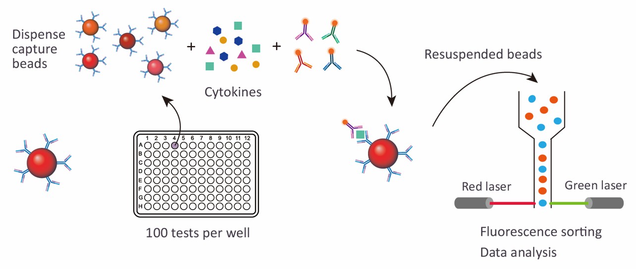
At Creative Proteomics, we utilize the advanced Luminex xMAP technology for our Human Apoptotic Cell Clearance 12-plex Panel analysis. This multiplexed assay platform allows for the simultaneous detection and quantification of multiple analytes from a single sample, enhancing throughput and reducing sample consumption.
Detection Method
Magnetic bead-based Luminex multiplex assay
Species
Human
Analytes Detected
| Species | Specification | Protein Targets | Applications | Price |
|---|---|---|---|---|
| Human | Human Apoptotic Cell Clearance 12-plex Panel | AXL, CD36, CALR (CRT), Gas6, LOX-1, MBL, Mer (MERTK), CD31 (PECAM-1), PAI-1 (Serpin), uPAR, RAGE, TYRO3 | Suitable for analyzing apoptotic cell clearance mechanisms related to inflammation, autoimmunity, and cancer progression. | +Inquiry |
Advantages of the Human Apoptotic Cell Clearance Luminex Assay
- High Sensitivity and Specificity: Luminex assays provide highly sensitive detection, capable of measuring low concentrations of analytes, which is crucial for accurately assessing apoptotic markers.
- Multiplexing Capability: The ability to analyze multiple targets simultaneously saves time and resources, facilitating comprehensive profiling of apoptotic cell clearance.
- Flexibility and Customization: Our services can be tailored to meet specific research needs, accommodating a variety of sample types and analyte profiles.
- Rapid Turnaround: With automated processes, we ensure a quick turnaround time for results, allowing for efficient progress in research timelines.

Sample Requirements for Human Apoptotic Cell Clearance Luminex Panel
| Sample Type | Volume Required | Storage Conditions | Stability |
|---|---|---|---|
| Whole Blood | 1-5 mL | 4°C (short term), -80°C (long term) | Up to 24 hours at 4°C, long-term at -80°C |
| Plasma | 0.5-1 mL | -80°C | Up to 6 months at -80°C |
| Serum | 0.5-1 mL | -80°C | Up to 6 months at -80°C |
| Cell Culture Supernatant | 0.5-1 mL | -80°C | Up to 6 months at -80°C |
| Synovial Fluid | 0.5-1 mL | -80°C | Up to 6 months at -80°C |
| Bronchoalveolar Lavage (BAL) | 0.5-1 mL | -80°C | Up to 6 months at -80°C |
| Urine | 5-10 mL | -80°C | Up to 6 months at -80°C |
| Tissue Homogenate | 0.5-1 g | -80°C | Up to 6 months at -80°C |
Application of Human Apoptotic Cell Clearance Panel
- Cancer Research: Understanding the dynamics of apoptotic cell clearance can reveal how tumors evade immune surveillance. The panel aids in identifying markers that indicate the efficiency of clearance in tumor microenvironments, helping to evaluate therapeutic responses and prognostic outcomes.
- Autoimmune Diseases: The panel is instrumental in studying conditions such as systemic lupus erythematosus (SLE) and rheumatoid arthritis, where impaired clearance of apoptotic cells may contribute to inflammation and autoimmunity. By analyzing the clearance mechanisms, researchers can identify potential therapeutic targets.
- Inflammatory Disorders: The panel can be used to investigate various inflammatory conditions, including chronic inflammatory diseases, by assessing the role of apoptotic cell clearance in modulating inflammation and tissue repair.
- Neurodegenerative Diseases: In diseases like Alzheimer's and Parkinson's, where neuronal cell death occurs, the panel helps in understanding how apoptotic cell clearance affects neuroinflammation and neurodegeneration, potentially revealing therapeutic strategies to enhance clearance mechanisms.
- Transplantation Biology: The panel can evaluate the role of apoptotic cell clearance in graft survival and rejection. Understanding how clearance mechanisms operate in transplanted tissues can improve strategies for managing transplant rejection and enhancing graft acceptance.
- Regenerative Medicine: By analyzing apoptotic cell clearance, researchers can optimize protocols in tissue engineering and regenerative medicine, as efficient clearance is vital for promoting tissue regeneration and repair.
- Biomarker Discovery: The panel aids in identifying novel biomarkers associated with effective or impaired apoptotic cell clearance, which can be used for diagnostics and monitoring therapeutic responses in various diseases.
- Pharmacodynamics: The panel is valuable for evaluating the effects of new drugs on apoptotic cell clearance, helping to predict drug efficacy and safety in preclinical and clinical studies.
In addition to preconfigured panels, we also offer customized analysis services. You can customize your own panel through our customization tool, or directly email us the targets you are interested in. A professional will contact you to discuss the feasibility of customization. We look forward to working with you!
| Protein Target | Description |
|---|---|
| AXL | A receptor tyrosine kinase involved in cell survival and proliferation; its activation is linked to tumor progression and resistance to apoptosis. |
| CD36 | A scavenger receptor that plays a role in lipid metabolism and immune response; associated with the clearance of apoptotic cells and regulation of inflammation. |
| CALR (CRT) | Calreticulin, a chaperone protein that is exposed on the surface of apoptotic cells, promoting their recognition and phagocytosis by immune cells. |
| Gas6 | A vitamin K-dependent protein that enhances the uptake of apoptotic cells by macrophages; involved in anti-inflammatory processes and tissue repair. |
| LOX-1 | A receptor for oxidized low-density lipoprotein (oxLDL); it plays a role in endothelial dysfunction and is implicated in the clearance of apoptotic cells. |
| MBL | Mannose-binding lectin, which activates the complement system; involved in innate immunity and recognition of apoptotic cells for clearance. |
| Mer (MERTK) | A receptor tyrosine kinase that mediates the engulfment of apoptotic cells by macrophages, playing a crucial role in maintaining tissue homeostasis. |
| CD31 (PECAM-1) | Platelet endothelial cell adhesion molecule; involved in cell adhesion and migration, and plays a role in the clearance of apoptotic cells in vascular contexts. |
| PAI-1 (Serpin) | Plasminogen activator inhibitor-1, which regulates fibrinolysis and is linked to tissue remodeling; elevated levels may affect apoptotic cell clearance. |
| uPAR | Urokinase-type plasminogen activator receptor, involved in cell signaling and migration; its expression is associated with tumor invasion and clearance processes. |
| RAGE | Receptor for advanced glycation end products; involved in the inflammatory response and clearance of apoptotic cells, with implications in various diseases. |
| TYRO3 | A member of the TAM receptor family, which plays a significant role in the phagocytosis of apoptotic cells and modulating immune responses. |
What is the significance of the "eat me" signals, such as phosphatidylserine exposure, in the context of apoptotic cell clearance?
During apoptosis, cells undergo specific morphological and biochemical changes, including the exposure of phosphatidylserine on their outer membrane. This "eat me" signal is crucial for the recognition and engulfment of apoptotic cells by phagocytes, primarily macrophages. This recognition helps ensure efficient clearance of dying cells, preventing the release of intracellular contents that could trigger inflammation and damage surrounding tissues. Understanding the mechanisms behind these signals can aid in developing therapies aimed at enhancing apoptotic cell clearance, particularly in conditions where this process is dysregulated, such as cancer and autoimmune diseases.
Can the Human Apoptotic Cell Clearance Panel be used to study the effects of therapeutic interventions on apoptotic cell clearance?
Yes, the Human Apoptotic Cell Clearance Panel is particularly useful for evaluating how various therapeutic interventions affect apoptotic cell clearance. By measuring changes in the levels of specific protein targets before and after treatment, researchers can gain insights into the effectiveness of therapies in modulating clearance mechanisms. This is especially relevant in cancer research, where treatments may influence the tumor microenvironment and the body's ability to clear apoptotic tumor cells, impacting overall treatment efficacy and patient outcomes.
What biomarkers associated with dysfunctional apoptotic cell clearance can be identified using the panel?
The Human Apoptotic Cell Clearance Panel detects several key protein targets, including AXL, Gas6, and Mer (MERTK), which are involved in the clearance process. Alterations in the levels of these proteins may serve as biomarkers for impaired clearance mechanisms. For instance, elevated levels of soluble AXL or decreased Mer activity can indicate a dysfunction in the clearance process, potentially contributing to chronic inflammation, autoimmunity, or tumor progression. Identifying these biomarkers can improve diagnostic accuracy and help tailor therapeutic strategies for affected patients.

