
- Services Overview
- FAQ
What is Human Angiogenesis?
Angiogenesis is the physiological process through which new blood vessels form from pre-existing ones, a critical component in growth, development, and wound healing. This complex process is orchestrated by a delicate balance of pro-angiogenic and anti-angiogenic factors that regulate the formation of new vasculature. Key molecules such as vascular endothelial growth factor (VEGF), fibroblast growth factor (FGF), and angiopoietins play pivotal roles in promoting angiogenesis. Conversely, inhibitors like thrombospondin-1 and endostatin serve to suppress unnecessary blood vessel formation. Disruption in this balance is often associated with pathological conditions, such as cancer, diabetic retinopathy, and rheumatoid arthritis, where angiogenesis may either be excessively stimulated or inadequately activated.
The analysis of human angiogenesis is fundamental for advancing our understanding of various biological processes. By analyze the key factors and pathways involved in angiogenesis, researchers and clinicians can:
- Uncover Mechanisms of Disease Development: Gain a deeper understanding of how abnormal angiogenesis contributes to various diseases, such as cancer, where excessive blood vessel formation supports tumor growth, or in ischemic conditions, where insufficient angiogenesis leads to tissue damage. This knowledge is essential for identifying potential points of intervention.
- Identify and Validate Biomarkers: Pinpoint specific biomarkers associated with angiogenic activity that can be used to monitor disease progression, assess the efficacy of therapeutic interventions, or serve as diagnostic indicators in research and clinical settings.
- Develop New Therapeutic Strategies: Inform the design of novel therapeutic approaches by targeting the critical molecules and pathways involved in angiogenesis. For example, researchers can develop inhibitors to suppress pathological angiogenesis or growth factors to promote vascularization in tissue engineering.
- Optimize Experimental Models: Enhance the accuracy and relevance of experimental models by integrating detailed knowledge of angiogenic processes. This can lead to more reliable predictions of how therapies will perform in clinical scenarios, ultimately reducing the time and cost associated with drug development.
Human Angiogenesis Panel at Creative Proteomics
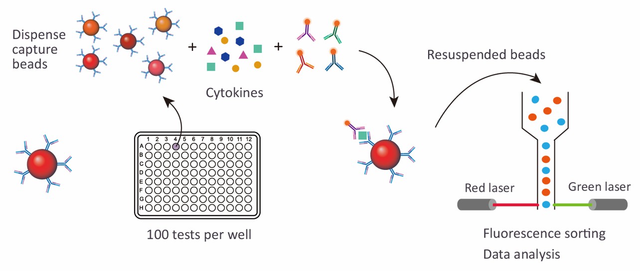
Creative Proteomics utilizes Luminex xMAP (Multi-Analyte Profiling) technology to perform Human Angiogenesis 20 Plex/39 Plex Panel analysis. This advanced multiplexing technology allows for the simultaneous quantification of multiple angiogenic factors from a single, small-volume sample. The Luminex xMAP system employs color-coded beads, each conjugated with a specific antibody, to capture target proteins. A laser-based detection system then identifies and quantifies these beads, providing robust and accurate measurements of up to 100 different analytes in a single reaction.
The use of Luminex xMAP technology by Creative Proteomics ensures high-throughput, highly sensitive, and precise analysis of angiogenic biomarkers. This technology is especially beneficial for:
- Simultaneous Detection: Ability to measure multiple biomarkers in parallel, reducing sample requirements and experimental variability.
- High Sensitivity and Specificity: Ensures that even low-abundance biomarkers are accurately quantified.
- Scalability: Suitable for both small-scale studies and large-scale clinical trials.
Detection Method
Magnetic bead-based Luminex multiplex assay
Species
Human
Analytes Detected
| Species | Specification | Protein Targets | Price |
|---|---|---|---|
| Human | Human Angiogenesis 20 Plex Panel | HMW Adiponectin/Acrp30, BMP-9, CD31/PECAM-1, CX3CL1/Fractalkine, DPPIV/CD26, HB-EGF, HGF, IGFBP-2, IL-10, Osteopontin/OPN, PDGF-AA, PIGF, Prolactin, Serpin F1/PEDF, Tenascin C, TIMP-1, uPA/Urokinase, VEGFR2/KDR/Flk-1, VEGFR3/FIt-4 | +Inquiry |
| Human | Human Angiogenesis 39 Plex Panel | Adiponectin/Acrp30, Angiogenin, Angiopoietin-1, Angiopoietin-2, Angiopoietin-like Protein 3/ANGPTL3, Angiopoietin-like Protein 4/ANGPTL4, Angiopoietin-like Protein 6/ANGPTL6, CCL2/JE/MCP-1, CCL3/MIP-1 alpha, CD117/c-kit, CXCL4/PF4, CXCL10/IP-10/CRG-2, CXCL16, EGF, Endoglin/CD105, Endostatin, ErbB2/Her2, ErbB3/Her3, FGF acidic/FGF1, FGF basic/FGF2, GDNF, GM-CSF, HGFR/C-MET, IGFBP-1, IGFBP-3, IL-1 beta/IL-1F2, IL-8/CXCL8, IL-6R alpha, Leptin/OB, MMP-3, MMP-8, MMP-9, Neuropilin-1, PDGF-BB, Pentraxin 3/TSG-14, Serpin E1/PAI-1, Thrombospondin-2, VEGF, VEGF-C | +Inquiry |
Advantages of the Human Angiogenesis Luminex Assay
- Multiplexing Capability: Unlike traditional ELISA, which can only measure one analyte at a time, the Luminex assay can analyze up to 100 analytes simultaneously. This multiplexing ability conserves sample volume and reduces assay time, allowing for a more efficient workflow.
- Enhanced Sensitivity: The Luminex assay is known for its high sensitivity, capable of detecting even minute quantities of biomarkers. This is particularly important in angiogenesis studies where certain key factors may be present at very low levels.
- Wide Dynamic Range: The assay provides a broad dynamic range, which allows for the accurate quantification of both high and low abundance targets in the same sample.
- Reproducibility: The robustness of the Luminex platform ensures consistent and reproducible results, which are critical for both research and clinical applications.
- Customizability: Researchers can customize the panel to include specific biomarkers of interest, enabling more targeted studies.
Sample Requirements for Human Angiogenesis Luminex Panel
| Sample Type | Volume Required | Storage Conditions | Special Instructions |
|---|---|---|---|
| Serum | 50 µL | -80°C | Collect using clot activator tubes; allow clotting for 30 min at room temperature, then centrifuge and aliquot. |
| Plasma (EDTA) | 50 µL | -80°C | Collect using EDTA tubes; centrifuge immediately after collection, aliquot, and freeze. |
| Cell Culture Supernatant | 500 µL | -80°C | Ensure cells are in logarithmic growth phase; collect supernatant after centrifugation to remove cells. |
| Tissue Lysate | 100 µg total protein | -80°C | Homogenize tissue in lysis buffer; centrifuge to remove debris and aliquot supernatant. |
Applications of Human Angiogenesis Panel Analysis
Cancer Research: The panel helps investigate the role of angiogenesis in tumor development and progression. By profiling angiogenic factors in cancer models, researchers can identify potential biomarkers for early detection, monitor treatment efficacy, and explore new therapeutic targets for anti-angiogenic strategies.
Cardiovascular Research: In cardiovascular studies, the panel is used to understand the mechanisms underlying ischemic diseases, such as heart disease and stroke. Analyzing angiogenic factors can reveal how blood vessel formation affects tissue repair and recovery, supporting the development of novel treatments to promote vascular regeneration.
Wound Healing and Tissue Engineering: The panel aids in evaluating the role of angiogenesis in wound healing and tissue regeneration. By assessing the factors that drive or inhibit angiogenesis, researchers can improve strategies for enhancing tissue repair and developing advanced biomaterials for regenerative medicine.
Ophthalmology: In the study of ocular diseases, such as diabetic retinopathy and age-related macular degeneration, the panel provides insights into the abnormal blood vessel growth that characterizes these conditions. This helps in developing targeted therapies to control or reverse pathological angiogenesis in the eye.
Inflammatory and Autoimmune Diseases: The panel is valuable for investigating the role of angiogenesis in chronic inflammatory and autoimmune conditions. By understanding how angiogenic factors contribute to disease pathology, researchers can identify potential targets for new anti-inflammatory treatments and interventions.
Biotechnology and Drug Development: The panel supports biotechnological innovation by providing critical data on angiogenic processes relevant to drug development and tissue engineering. It helps in screening and validating new drugs that affect angiogenesis, as well as in designing more effective biotechnological products and therapeutic approaches.
In addition to preconfigured panels, we also offer customized analysis services. You can customize your own panel through our customization tool, or directly email us the targets you are interested in. A professional will contact you to discuss the feasibility of customization. We look forward to working with you!
How should I collect and prepare samples to ensure the best results?
Serum: Use clot activator tubes for collection, allow the blood to clot for 30 minutes at room temperature, then centrifuge at 1,000-2,000 x g for 10 minutes. Aliquot the serum into clean tubes and store at -80°C.
Plasma (EDTA): Collect blood in EDTA tubes, centrifuge within 30 minutes at 1,000-2,000 x g for 10 minutes, then aliquot and freeze at -80°C immediately.
Cell Culture Supernatant: Harvest supernatants from cells in the logarithmic growth phase, centrifuge at 300 x g for 10 minutes to remove cells, aliquot the supernatant, and store at -80°C.
Tissue Lysate: Homogenize tissue in cold lysis buffer with protease inhibitors, centrifuge at 10,000 x g for 15 minutes to clear debris, then aliquot the supernatant and store at -80°C.
How can I prevent sample degradation during storage and transport?
To prevent sample degradation:
- Store all samples at -80°C as soon as possible after preparation.
- Avoid repeated freeze-thaw cycles by aliquoting samples before storage.
- Use dry ice for transport to maintain low temperatures.
- Ensure samples are properly sealed and labeled to prevent contamination and misidentification.
What factors could affect the accuracy of my assay results?
Improper sample handling: Delayed processing, repeated freeze-thaw cycles, or incorrect storage temperatures can degrade biomolecules.
Contamination: Contaminants introduced during sample collection or processing can interfere with assay detection.
Sample volume: Insufficient sample volume can lead to inadequate detection of biomarkers, affecting the reliability of results.
Assay interference: Substances such as hemoglobin, lipids, or certain drugs in the sample may interfere with the assay.
What controls should I include in my experiment?
To ensure data reliability, include the following controls:
- Positive Control: A known sample with established levels of angiogenic factors to validate the assay's performance.
- Negative Control: A sample known to lack angiogenic activity, ensuring the assay does not produce false positives.
- Blank Control: Buffer or media used in sample preparation without any biological sample, to check for contamination or non-specific binding.
- Replicates: Perform technical replicates of each sample to assess the consistency of the results.
Can I use frozen samples, and how should they be handled before analysis?
Yes, frozen samples can be used. Prior to analysis:
- Thaw samples slowly on ice to minimize degradation.
- Gently mix after thawing to ensure homogeneity.
- Avoid multiple freeze-thaw cycles, which can degrade sensitive biomolecules and affect the accuracy of the assay.
How can I ensure my sample concentration is within the assay's dynamic range?
To ensure sample concentration falls within the assay's dynamic range:
- Dilute samples systematically and perform a pilot test to identify the optimal dilution factor.
- Consult the assay's documentation for recommended concentration ranges of specific analytes.
- Include a standard curve with known concentrations of the target analytes to accurately quantify sample concentrations.
What should I do if I encounter unexpected results or low signal in my assay?
If you encounter unexpected results or low signal:
- Review sample preparation protocols: Ensure that all steps were followed correctly, and samples were stored properly.
- Check for interference: Investigate whether any substances in your samples might be interfering with the assay.
- Re-run controls: Ensure that controls are performing as expected. Poor performance in controls may indicate issues with the assay itself.
- Consult technical support: If issues persist, contact Creative Proteomics for troubleshooting assistance or to discuss the possibility of assay optimization.
How much sample volume should I prepare if I need to run multiple tests?
To run multiple tests, ensure that you prepare sufficient sample volume to accommodate the number of assays planned. For instance:
- If the assay requires 50 µL per test, and you plan to run triplicates, prepare at least 200 µL to account for pipetting variations and any potential repeats.
- Always prepare a bit more than the calculated amount to avoid running out of sample.
Can the assay be adapted for high-throughput screening?
Yes, the Human Angiogenesis Luminex Assay is well-suited for high-throughput screening. It allows for the simultaneous analysis of multiple biomarkers across a large number of samples, making it ideal for screening applications. Ensure that your laboratory setup includes automated pipetting systems and appropriate plate readers to fully leverage the assay's high-throughput capabilities.