- Services Overview
- Analytes Details
- FAQ
What is Mouse Inflammatory Factor?
Inflammation is a key biological response to infection, injury, or tissue damage, crucial for immune defense and tissue repair. In mice, as in humans, inflammation is mediated by a variety of molecules, including cytokines, chemokines, and growth factors, which regulate immune cell activity and recruitment. While inflammation is essential for defense, its dysregulation can lead to chronic inflammation, contributing to diseases such as autoimmune disorders, cancer, and cardiovascular conditions.
Key Inflammatory Factors
- Cytokines: Small proteins that mediate immune responses. Key proinflammatory cytokines include TNF-Alpha, IL-1, IL-6, and IFN-Gamma, which initiate and sustain inflammation.
- Chemokines: Molecules like MCP-1, MIP-1, and RANTES control immune cell migration to sites of inflammation.
- Growth Factors: Molecules such as IL-12p70, IL-15, and IL-13 regulate inflammation and tissue repair.
Mice are widely used in inflammation research due to their genetic similarities to humans. Mouse models help study the mechanisms of inflammatory diseases, including autoimmune diseases, cancer, infections, and chronic inflammation. Monitoring inflammatory factors in mice provides insights into disease mechanisms and the efficacy of potential treatments.
Mouse Inflammatory Panel at Creative Proteomics
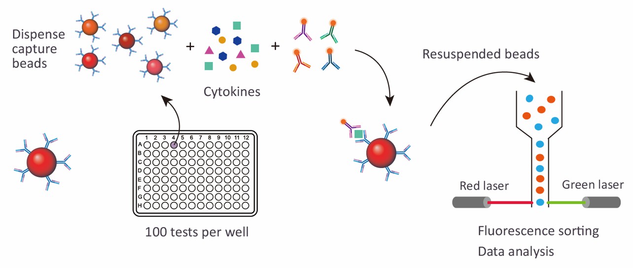
At Creative Proteomics, we employ Luminex xMAP technology to perform our Mouse Inflammatory 15-plex Panel assays. Luminex xMAP (Multi-Analyte Profiling) is a powerful technology that allows simultaneous detection and quantification of multiple analytes in a single sample. Using a suspension array format, xMAP technology provides researchers with a high degree of multiplexing capability.
Our mouse inflammatory panel utilizes bead-based assays, with each bead type coated with a specific capture antibody to detect a distinct inflammatory factor. When a sample is added to the array, analytes present in the sample bind to their corresponding capture antibodies, and subsequent detection occurs using fluorescently-labeled secondary antibodies. This allows for the simultaneous measurement of multiple inflammatory markers in a single well, drastically reducing both sample and reagent consumption.
Detection Method
Magnetic bead-based Luminex multiplex assay
Species
Mouse
Analytes Detected
| Species | Specification | Protein Targets | Applications | Price |
|---|---|---|---|---|
| Mouse | Mouse Inflammatory 15-plex Panel | BAFF/BLyS/TNFSF13B, CCL5/RANTES, Chitinase 3-like 1, IFN-gamma, IL-2, IL-6, IL-10, IL-12 p70, IL-27, IL-6R alpha, MMP-2, MMP-3, TNF-alpha, TNF RI/TNFRSF1B, TWEAK/TNFSF12 | Suitable for analyzing inflammatory profiles in autoimmune diseases, cancer, infections, and chronic inflammatory conditions. | +Inquiry |
Advantages of the Mouse Inflammatory Luminex Assay
- High Multiplexing Capability: The Luminex xMAP technology enables the simultaneous detection of multiple inflammatory markers in a single sample. This reduces assay time and resource consumption while providing a comprehensive analysis of inflammatory responses.
- Superior Sensitivity and Precision: The assay offers exceptional sensitivity with detection limits as low as <1 pg/mL for certain analytes. This allows for accurate detection of low-concentration inflammatory factors, crucial for studying early-stage inflammation or subtle immune responses.
- Cost and Sample Efficiency: By analyzing multiple inflammatory markers in one test, the Luminex assay minimizes both sample and reagent usage, making it a cost-effective solution for high-throughput studies.
- Wide Range of Analytes: The mouse inflammatory Luminex panel includes a broad spectrum of cytokines, chemokines, and growth factors, enabling a thorough profile of the inflammatory environment in mouse models.
- Low Sample Volume Requirement: Each test requires just 25 µL of sample, making it suitable for studies with limited sample availability, such as those involving rare tissues or small animals.
- Customizable Panels: The flexibility to customize the panel allows researchers to focus on specific analytes, tailoring the assay to meet unique experimental needs.
- Reproducible and Reliable Results: The Luminex technology delivers consistent, high-quality data with low inter-assay variability, ensuring that results are reproducible across different experiments.

Sample Requirements for Mouse Inflammatory Luminex Panel
| Sample Type | Required Volume | Sample Preparation | Storage Conditions | Notes |
|---|---|---|---|---|
| Serum | 25 µL per test | Collect by centrifugation after blood clotting. Avoid hemolysis. | Store at -80°C for long-term, or 4°C for short-term. | Ensure the sample is free of particulate matter. |
| Plasma | 25 µL per test | Collect using EDTA or heparin tubes. Centrifuge at 1,500 x g for 10 minutes. | Store at -80°C for long-term, or 4°C for short-term. | Do not use samples containing clots. |
| Cell Culture Supernatant | 25 µL per test | Centrifuge to remove any cellular debris. | Store at -80°C for long-term, or 4°C for short-term. | Ensure no contamination with serum or plasma. |
| Bodily Fluids (e.g., saliva, urine) | 25 µL per test | Collect using clean containers. Centrifuge to remove particles if needed. | Store at -80°C for long-term, or 4°C for short-term. | If sample volume is limited, perform concentration steps. |
| Tissue or Cell Lysate | 25 µL per test | Homogenize tissue in lysis buffer, centrifuge to remove debris. | Store at -80°C for long-term, or 4°C for short-term. | Use proper protease inhibitors during sample preparation. |
Application of Mouse Inflammatory Panel
- Autoimmune diseases
Autoimmune diseases like rheumatoid arthritis (RA) and lupus are characterized by chronic inflammation. The mouse inflammatory factor Luminex panel can detect key cytokines such as TNF-alpha, IL-6, and IL-1β, which are typically elevated in these conditions. For example, in a collagen-induced arthritis mouse model (a model for RA), researchers used the panel to monitor the progression of inflammatory markers. Elevated levels of IL-6 and TNF-alpha were found to correlate with disease severity, guiding the development of therapies that target these cytokines. The data helped evaluate the efficacy of biologics like TNF inhibitors and IL-6 receptor blockers, which are currently used to treat RA.
- Cancer research
Inflammation is a key driver of cancer progression, particularly in the tumor microenvironment. The mouse inflammatory factor Luminex panel is used to study the role of cytokines like TNF-alpha, IL-6, and MCP-1 in promoting tumor growth, metastasis, and immune evasion. In a mouse model of breast cancer, the panel was used to monitor inflammatory cytokines in tumors. Elevated levels of IL-6 and TNF-alpha were associated with increased tumor growth and poor prognosis. This data helped inform the development of targeted therapies aimed at modulating these inflammatory pathways to slow down cancer progression.
- Infectious diseases
The immune response to infection is driven by complex networks of cytokines. The mouse inflammatory factor Luminex panel is commonly used to monitor these immune responses during infection. For example, in a mouse model of influenza, the panel was used to track the levels of IFN-gamma, IL-6, and MIP-1α in the lungs of infected mice. High levels of IFN-gamma were correlated with effective antiviral immunity, while IL-6 and MIP-1α were involved in recruiting immune cells to the site of infection. This data is critical for evaluating the efficacy of antiviral drugs and for understanding how inflammation affects immune defense against pathogens.
- Neuroinflammation and neurodegenerative diseases
Neuroinflammation is implicated in a range of neurodegenerative diseases such as Alzheimer's disease, Parkinson's disease, and multiple sclerosis. The mouse inflammatory factor Luminex panel is used to investigate how inflammatory cytokines like TNF-alpha, IL-1β, and IL-6 contribute to neurodegeneration. In a mouse model of Alzheimer's disease, researchers used the panel to measure levels of these cytokines in the brain. Elevated levels of IL-6 and TNF-alpha were associated with cognitive decline and amyloid plaque formation. This data informed therapeutic approaches aimed at reducing neuroinflammation, such as using TNF-alpha inhibitors or IL-1β antagonists, which have shown promise in preclinical studies.
- Cardiovascular diseases
Chronic inflammation plays a central role in cardiovascular diseases such as atherosclerosis, heart failure, and myocardial infarction. The mouse inflammatory factor Luminex panel is valuable for studying inflammatory markers like IL-6, MCP-1, and TNF-alpha in cardiovascular disease models. For example, in a mouse model of atherosclerosis, researchers used the panel to track cytokine levels in the blood. Elevated levels of TNF-alpha and IL-6 were linked to increased plaque formation and vascular inflammation. This data helped assess the potential of anti-inflammatory therapies, such as statins and IL-1β inhibitors, to reduce inflammation and prevent the progression of cardiovascular disease.
- Injury and tissue repair
Inflammation is a critical component of tissue repair following injury. The mouse inflammatory factor Luminex panel can be used to monitor cytokines involved in tissue healing and fibrosis. In a mouse liver fibrosis model, researchers used the panel to measure cytokines like IL-13, TGF-β, and MMP-2, which are involved in fibrosis. Elevated levels of IL-6 and MCP-1 were found to be associated with early stages of tissue repair, while TGF-β played a key role in fibrosis. By targeting these cytokines with specific inhibitors, researchers were able to reduce liver fibrosis and improve tissue regeneration, demonstrating the potential for cytokine modulation in regenerative medicine.
- Drug development and toxicology
The mouse inflammatory factor Luminex panel is a valuable tool for evaluating the impact of new drugs or toxic compounds on inflammation. In a toxicity study of a novel chemotherapeutic agent, the panel was used to measure inflammatory markers such as IL-6, TNF-alpha, and IL-10 to assess the immune response. If elevated levels of these cytokines were observed, it could indicate that the drug is inducing unwanted inflammation, which may contribute to adverse side effects. This data helps refine drug formulations to improve their safety profiles and avoid unwanted inflammatory responses during treatment.
In addition to preconfigured panels, we also offer customized analysis services. You can customize your own panel through our customization tool, or directly email us the targets you are interested in. A professional will contact you to discuss the feasibility of customization. We look forward to working with you!
| Protein Target | Description |
|---|---|
| BAFF/BLyS/TNFSF13B | A cytokine essential for B cell survival and maturation. It plays a critical role in humoral immunity and is involved in autoimmune diseases such as lupus, rheumatoid arthritis, and Sjögren's syndrome. |
| CCL5/RANTES | A chemokine that promotes the recruitment and activation of immune cells, particularly T cells and eosinophils, to sites of inflammation. Elevated levels of CCL5 are associated with inflammatory conditions such as asthma, rheumatoid arthritis, and atherosclerosis. |
| Chitinase 3-like 1 | A glycoprotein involved in inflammation and tissue repair. It is elevated in diseases with chronic inflammation, such as asthma, rheumatoid arthritis, and cancer. It is also implicated in fibrosis and tissue remodeling. |
| IFN-gamma | A pro-inflammatory cytokine produced by Th1 cells, NK cells, and CD8+ T cells. It is essential for defense against intracellular pathogens, activates macrophages, and modulates adaptive immune responses. Elevated levels are seen in autoimmune disorders and infections. |
| IL-2 | A cytokine crucial for T cell proliferation and differentiation. It promotes the growth and survival of T cells and plays a key role in immune responses, particularly in the activation of CD4+ and CD8+ T cells. It is important for both adaptive immunity and immune tolerance. |
| IL-6 | A pleiotropic cytokine involved in immune responses, inflammation, and metabolic processes. It is produced during acute-phase responses and is associated with diseases such as rheumatoid arthritis, sepsis, and various cancers. |
| IL-10 | An anti-inflammatory cytokine that regulates immune responses by inhibiting pro-inflammatory cytokine production. It plays a critical role in immune tolerance and limiting excessive tissue damage during inflammation. |
| IL-12 p70 | A cytokine that stimulates the production of IFN-gamma and promotes Th1 differentiation. It is important in the immune defense against intracellular pathogens and has a role in autoimmune inflammation. |
| IL-27 | A cytokine involved in immune regulation, promoting Th1 differentiation and influencing the balance between pro-inflammatory and regulatory immune responses. It plays a role in autoimmune diseases and chronic inflammation. |
| IL-6R alpha | The receptor for IL-6, involved in mediating the effects of IL-6. It is crucial in regulating immune responses and inflammation and is a target for therapies aimed at treating autoimmune diseases and cancers. |
| MMP-2 | A matrix metalloproteinase involved in the degradation of extracellular matrix proteins, essential for tissue remodeling, wound healing, and angiogenesis. It is implicated in cancer metastasis and inflammatory diseases. |
| MMP-3 | Another matrix metalloproteinase that degrades extracellular matrix components and is involved in tissue remodeling, inflammation, and tissue repair. It is elevated in diseases such as rheumatoid arthritis and osteoarthritis. |
| TNF-alpha | A potent pro-inflammatory cytokine that regulates immune responses and inflammation. It plays a central role in diseases such as rheumatoid arthritis, inflammatory bowel disease, and sepsis. |
| TNF RI/TNFRSF1B | A receptor for TNF-alpha, involved in mediating TNF signaling. It plays a key role in regulating immune responses, inflammation, and apoptosis, and is a target for therapies aimed at modulating TNF-mediated diseases. |
| TWEAK/TNFSF12 | A cytokine that regulates tissue repair, immune responses, and inflammation. It plays a role in various diseases, including autoimmune diseases, cancer, and cardiovascular conditions. It is involved in the balance between inflammation and tissue regeneration. |
Can I customize the Mouse Inflammatory Panel to include specific cytokines or analytes?
Yes, we offer customizable panels based on the specific needs of your research. While our standard mouse inflammatory panel includes 15 key cytokines, chemokines, and growth factors, we can tailor the panel to focus on specific analytes relevant to your research. This flexibility allows you to add or remove factors based on your experimental goals, making it an ideal solution for a variety of disease models, such as autoimmune disorders, cancer, or infections.
How does the Luminex xMAP technology compare to other methods in terms of accuracy and throughput?
Luminex xMAP technology is highly accurate and enables multiplex detection of up to 100 analytes simultaneously in a single sample. This significantly reduces the time and resources required for traditional methods, such as ELISA, which would require individual assays for each analyte. The high sensitivity of Luminex technology, coupled with its ability to detect multiple analytes at once, makes it an ideal solution for large-scale, high-throughput studies, especially when dealing with limited sample volumes. Moreover, the technology ensures low inter-assay variability, delivering reliable and reproducible results across different experiments.
What is the cost of using the mouse inflammatory panel?
The pricing for the mouse inflammatory panel varies based on the specific requirements of your study, including the number of analytes tested and the number of samples to be processed. For exact pricing, we recommend contacting our sales team to get a customized quote based on your project needs. In addition, we offer bulk discounts for large-scale studies and consultations to ensure that the panel is optimally configured for your research goals.

