
AMPK (Adenosine 5'-monophosphate (AMP)-activated protein kinase), as an important kinase regulating energy homeostasis, is one of the central regulators of eukaryotic cell and organism metabolism. It is responsible for regulating cellular capacity input and output and maintaining the smooth functioning of cellular physiological activities. AMPK is also a key protein involved in various signaling pathways.
AMPK Structure
There is no complete structure of AMPK heterotrimer, but the structure of each structural domain has been determined by X-ray crystallography. The crystallographic structure consists mainly of the carboxy-terminal structural domain of the β-subunit (β-CTD), the C-terminal structural domain of the α-subunit (α-CTD), and the CBS structural domain of the γ-subunit.
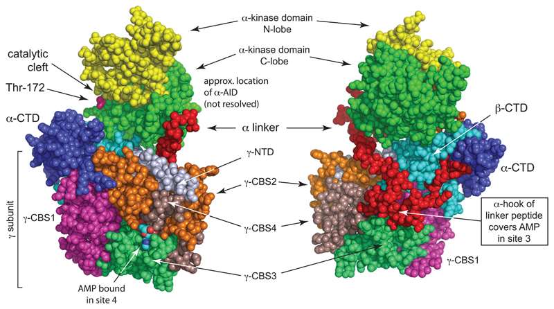 Crystal structure of mammalian AMPK (Hardie et al., 2012)
The alpha subunit has a catalytic region at the N-terminal end of the serine/threonine protein kinase. The intermediate self-inhibitory region contains a variable binding site on the C-terminal subunit binding region, which can be involved in AMP binding.
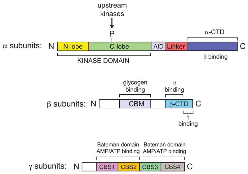 Domain map of typical mammalian AMPK (Hardie et al., 2012)
The α-subunit is primarily responsible for catalysis, where phosphorylation of the threonine 172 (Thr172) site is required for AMPK activation.
The β-subunit is often regarded as a scaffold for linking the α and γ subunits. However, as we have learned more about it, researchers have found that the β subunit can also be involved in the functional expression of AMPK by regulating the phosphorylation status and activity of the AMPK complex.
The γ-subunit contains four cystathione-β-synthase structural domains. When the four structural domains are linked in series, they form the Bateman domain. When the Bateman domain is altered, it affects the ability of AMPK to bind AMP, which in turn affects cellular energy metabolism.
AMPK Signaling Pathway
Numerous growth factors, altered cellular stress, altered cell motility, and altered energy uptake can all activate AMPK upstream kinases.
LKB1, CaMKK, and PKA are the main upstream kinases that activate AMPK, and their activation phosphorylates T172 of AMPK.
AMPK can further phosphorylate a variety of downstream transcription factors involved in the regulation of glucose metabolism, lipid metabolism, cell proliferation and other processes.
Functions of AMPK
Once activated, AMPK regulates four major classes of metabolism in mammals: protein metabolism, lipid metabolism, sugar metabolism, and autophagy and mitochondrial homeostasis, encompassing almost the entire physiological metabolic activity of living organisms. AMPK can stop the metabolism of rapid tumor proliferation as well as restore the normal function of liver and other tissues in diabetic patients is a hot topic in disease research.
- AMPK and viral infections
The pharmacological activity of AMPK plays an important role in the accumulation of lipids in the cells and in the self-replication of the virus.
HCV infection increases the level of reactive oxygen species in host cells, alters the NAD/NADH ratio and reduces the levels of silent information regulator 1 (SIRT1) and AMPK, thereby altering hepatocyte metabolism, a mechanism associated with the development of metabolic diseases of the liver.
Human cytomegalovirus (HCMV) activates AMPK in host cells via CaMKK, which increases the transport of glucose by GLUT4, thereby increasing host cell replication. Compound C, an AMPK inhibitor, blocks the HCMV-induced glycolysis process, thereby attenuating HCMV replication.
- AMPK and bacterial infections
After parasitizing the host, pathogenic bacteria modulate the AMPK signaling pathway to alter the host metabolic environment and compete with the host for the necessary energy to meet their own growth and developmental requirements.
Early infection with Salmonella typhimurium can activate AMPK. As infection progresses, S. typhimurium forces the degradation of the Sirt1/LKB1/AMPK complex in the lysosome, which limits AMPK activity and leads to mTOR activation, thereby inhibiting host cell autophagy in favor of S. typhimurium multiplication.
- AMPK and parasitic infections
Parasites need to parasitize their hosts and obtain nutrients such as nitrogen, amino acids, ribose, galactose and glucose from the host environment to facilitate their survival and reproduction. Therefore, the dynamic interaction of energy between host and parasite is necessary to effectively coordinate the different developmental stages of the parasite, and AMPK plays an important role in this process.
ω3-PUFAs activate the AMPK signaling pathway via the upstream kinase CaMKKβ, which induces autophagy in Toxoplasma gondii-infected macrophages, thereby limiting the growth and reproduction of intracellular Toxoplasma.
Creative Proteomics develops AMPK signaling pathway protein target detection platform based on luminex multiplex technology. Contact us to learn more.
Reference:
- Hardie, D. G., Ross, F. A., & Hawley, S. A. (2012). AMPK: a nutrient and energy sensor that maintains energy homeostasis. Nature reviews Molecular cell biology, 13(4), 251-262.