
Standard Luminex panels cover the most commonly requested cytokines for human and mouse — but they cannot anticipate every experimental need. When a project requires CXCL9 and CXCL10 alongside a standard 8-plex, or when working with guinea pig, rabbit, or porcine models where off-the-shelf options are thin, a custom-designed panel becomes the only viable path.
Custom Luminex design is not simply a matter of mixing bead regions from different kits — it requires systematic antibody pair screening, cross-reactivity validation across every bead-analyte combination, and panel-level QC that ensures the assembled multiplex performs as a unified assay rather than a collection of individual tests. For an initial feasibility check, use our Luminex Multiplex Assay Customization Tool.
The Custom Panel Design Workflow
| Phase | What Happens | Typical Duration |
|---|---|---|
| 1. Analyte selection & feasibility | Target analytes checked against antibody catalogue. Unavailable analytes flagged for custom sourcing or cross-reactive screening. | 1–2 weeks |
| 2. Cross-reactivity screening | Each antibody-bead pair tested against every other analyte. Pairs producing >1% cross-signal flagged for replacement or panel splitting. | 3–4 weeks |
| 3. Panel assembly & standard curve QC | Validated bead regions combined into single multiplex. 7-point standard curves + blank, triplicate across 3 plates. Linearity, LLOQ, and inter-plate precision verified per analyte. | 2–3 weeks |
| 4. Matrix validation | Spike-recovery (80–120%), dilution linearity (parallelism: slope ratio 0.9–1.1), and endogenous QC testing in intended matrix. | 2 weeks |
| 5. Pilot run | 3–5 representative study samples per group tested to confirm biologically interpretable data. | 1–2 weeks |
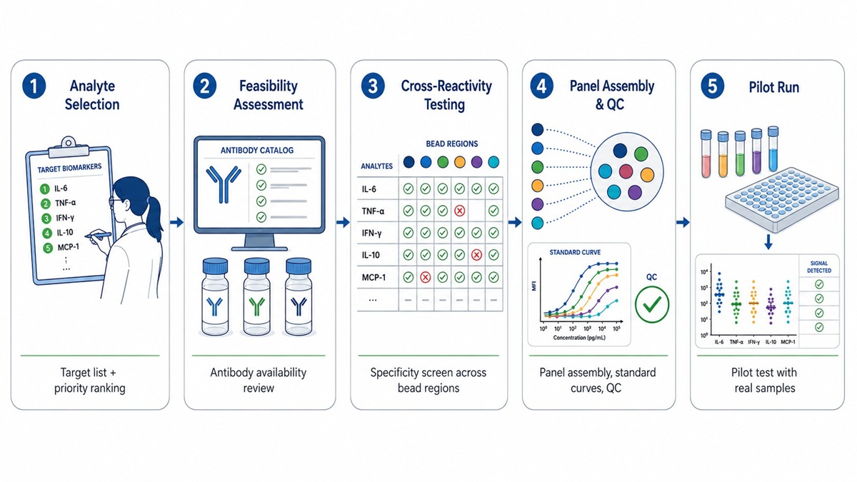 Five-phase workflow for custom Luminex panel development: analyte selection, cross-reactivity screening, panel assembly, matrix validation, and pilot testing.
Cross-Reactivity Validation: The Technical Core
In a multiplex Luminex assay, each bead region is coated with a capture antibody specific to one analyte. A biotinylated detection antibody binds the captured target and generates signal via streptavidin-PE. The risk: a detection antibody intended for analyte A may cross-recognise analyte B when B is captured on its own bead region, producing false signal at bead address A.
The systematic approach
- Prepare individual high-concentration standards (50–100 ng/mL) for each analyte.
- Run each single-analyte standard against every bead region in the proposed multiplex. For a 15-plex: 15 × 15 = 225 measurements.
- Calculate percentage cross-signal: (signal from analyte B on bead A / signal from analyte A on bead A) × 100.
- The industry-standard threshold for non-cross-reactivity is <1%. 1–5% is borderline; >5% is unacceptable.
Resolving cross-reactivity
- Option 1: Replace the problematic detection antibody with an alternative clone.
- Option 2: Move one analyte to a separate, parallel panel — run Panel A and Panel B on separate sample aliquots.
- Option 3: If biological concentrations of the two analytes always differ by >100×, accept documented cross-reactivity with caveats.
The N² scaling problem
The validation burden scales quadratically: 10-plex = 100 measurements, 20-plex = 400, 30-plex = 900. Adding a single analyte to an existing panel that was not validated together means re-running the full cross-reactivity matrix, not just testing the new analyte against existing ones.
Species-Specific Antibody Availability
| Species | Validated Analytes | Commonly Available Markers |
|---|---|---|
| Human | 200+ | IL-1β, IL-2, IL-4, IL-5, IL-6, IL-8, IL-10, IL-12p70, IL-13, IL-17A, IFN-γ, TNF-α, GM-CSF, MCP-1, MIP-1α/β, IP-10, VEGF, and many more |
| Mouse | 150+ | IL-1β, IL-2, IL-4, IL-5, IL-6, IL-10, IL-12p70, IL-13, IL-17A, IFN-γ, TNF-α, GM-CSF, KC, MCP-1, MIP-1α, RANTES, and many more |
| Rat | 40+ | IL-1β, IL-2, IL-4, IL-6, IL-10, IL-12p70, IL-13, IL-17A, IFN-γ, TNF-α, GM-CSF, MCP-1, VEGF |
| Porcine | 13–19 | GM-CSF, IFN-γ, IL-1α, IL-1β, IL-1Ra, IL-2, IL-4, IL-6, IL-8, IL-10, IL-12p40, IL-18, TNF-α |
| Rabbit | ~15 | IFN-γ, IL-1β, IL-2, IL-4, IL-6, IL-8, IL-10, IL-12, IL-17A, TNF-α, GM-CSF, MCP-1 |
| Canine | ~15 | IL-2, IL-4, IL-6, IL-8, IL-10, IFN-γ, TNF-α, MCP-1, GM-CSF, IL-12p40 |
| Guinea Pig | ≤10 | IFN-γ, IL-1β, IL-4, IL-6, IL-10, TNF-α |
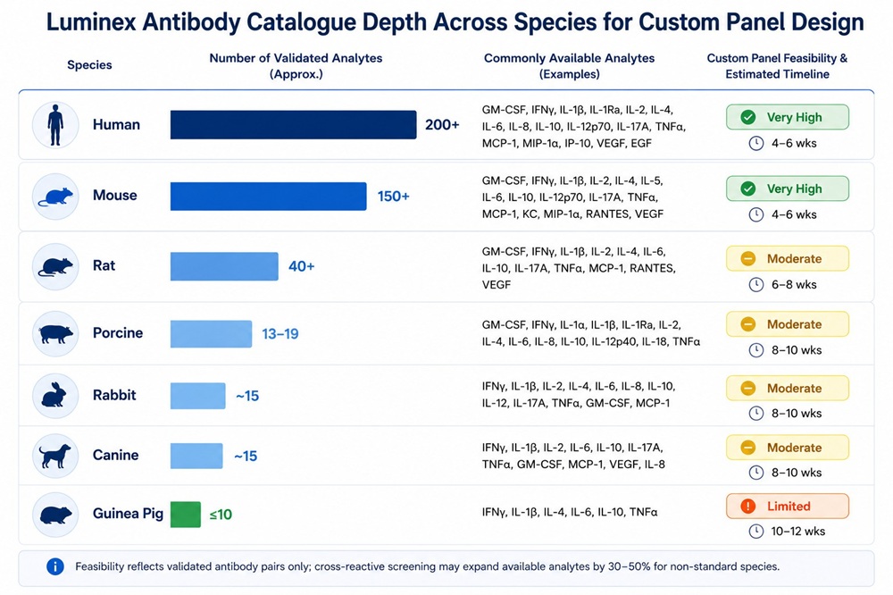 Luminex analyte availability by species, showing the antibody catalogue depth and custom panel feasibility from human (200+ analytes) to guinea pig (≤10).
Standard Curve Validation in Custom Panels
A standard curve validated for single-plex may not perform identically when placed into multiplex. For each analyte in a custom panel, the standard curve is reconstructed on the assembled multiplex — not carried over from the original single-plex data.
- Format: 7-point series + blank, triplicate across three independent plates.
- Curve fit: 5-parameter logistic (5PL) weighted (1/y²) preferred; 4PL acceptable if asymmetry tests fail.
- Back-calculation: Each standard point within ±20% of nominal (±25% at LLOQ and ULOQ).
- LLOQ: Lowest standard with recovery 80–120% and %CV ≤ 25%.
- Dynamic range: 3–4 orders of magnitude per analyte.
- Inter-plate precision: %CV ≤ 20% across plates for each standard point.
When to Split a Custom Panel into Two
- Persistent cross-reactivity: Two analytes consistently produce >1% cross-signal after testing alternative antibody clones.
- Incompatible concentration ranges: One analyte requires dilution while another requires concentrated sample — and this cannot be reconciled with a single protocol.
- Different preparation requirements: One analyte requires acid activation (e.g., TGF-β1) while others do not.
Running Panel A (8-plex) and Panel B (6-plex) on separate aliquots from the same sample is a standard resolution. Sample volume approximately doubles, but data quality is preserved.
Common Custom Panel Scenarios
- Th17 pathway: IL-17A, IL-17F, IL-21, IL-22, IL-23, IL-6, TGF-β1, TNF-α — typically distributed across 3–4 different standard panels.
- Angiogenesis: VEGF-A, VEGF-C, VEGF-D, FGF-2, PDGF-BB, HGF, angiopoietin-2, endostatin — concentrated into one custom panel from 4–5 standard kits.
- Chemokine-focused: CXCL9 (MIG), CXCL10 (IP-10), CXCL11 (I-TAC), CCL2 (MCP-1), CCL3 (MIP-1α), CCL5 (RANTES), CCL11 (Eotaxin) — chemokines are under-represented in standard inflammation panels.
For consultation on panel design, see our Luminex cytokine detection service.
What to Include in a Custom Panel Request
- Analyte list with official gene symbols: Use CXCL8 (not IL-8), CCL2 (not MCP-1). Mark essential vs optional.
- Species and strain: Matters for cross-reactive antibody feasibility.
- Sample matrix and anticoagulant: EDTA-plasma, heparin-plasma, citrate-plasma, serum, supernatant, tissue lysate, BALF, saliva, or other.
- Sample volume available: 25 µL per well standard; submit 2–4× for repeats and dilutions.
- Expected concentration ranges: Determines standard curve span.
- Study design: Groups, samples per group, replicates.
References:
- Vignali, D. A. A. "Multiplexed particle-based flow cytometric assays." Journal of Immunological Methods 243.1–2 (2000): 243–255. DOI: 10.1016/S0022-1759(00)00238-6
- de Jager, W., et al. "Improved multiplex immunoassay performance in human plasma and synovial fluid following removal of interfering heterophilic antibodies." Journal of Immunological Methods 300.1–2 (2005): 124–135. DOI: 10.1016/j.jim.2005.03.009
- Tighe, P. J., et al. "ELISA in the multiplex era: Potentials and pitfalls." Proteomics – Clinical Applications 9.3–4 (2015): 406–422. DOI: 10.1002/prca.201400130
- Chowdhury, F., et al. "Validation and comparison of two multiplex technologies, Luminex and Mesoscale Discovery, for human cytokine profiling." Journal of Immunological Methods 340.1 (2009): 55–64. DOI: 10.1016/j.jim.2008.10.007
- Leng, S. X., et al. "ELISA and multiplex technologies for cytokine measurement in inflammation and aging research." Journal of Gerontology: Medical Sciences 63.8 (2008): 879–884. DOI: 10.1093/gerona/63.8.879