What is PAI-1 Signaling Pathway?
Plasminogen Activator Inhibitor-1 (PAI-1) is a crucial protein in the human body, playing a central role in various physiological and pathological processes as a potent protease inhibitor. Within cells, PAI-1 primarily inhibits tissue plasminogen activator (tPA) and urokinase plasminogen activator (uPA), enzymes responsible for converting plasminogen into plasmin—an essential enzyme for breaking down fibrin blood clots and other extracellular matrix components. By inhibiting tPA and uPA, PAI-1 effectively regulates the fibrinolytic system.
The significance of the PAI-1 signaling pathway lies in maintaining the balance between clot formation (thrombosis) and clot dissolution (fibrinolysis). Elevated PAI-1 levels reduce fibrinolysis, increasing the risk of excessive blood clot formation in conditions like thrombotic disorders. Conversely, deficiencies or reduced PAI-1 expression lead to hyper-fibrinolysis, raising the risk of bleeding.
This pathway is a critical regulatory mechanism within cells, with PAI-1 at its core functioning as a protease inhibitor. It impacts various physiological and pathological processes, such as hemostasis and thrombosis. Additionally, PAI-1 plays a pivotal role in cellular migration and tissue remodeling by modulating proteolysis, affecting processes like wound healing and cancer metastasis.
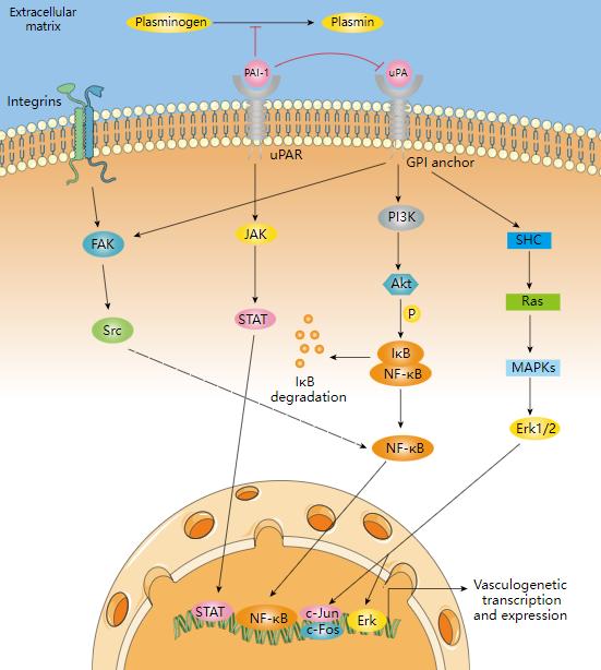 Plasminogen activator inhibitor-1 signaling pathway.
Regulation of the PAI-1 Signaling Pathway
Understanding the intricate regulation of the PAI-1 (Plasminogen Activator Inhibitor-1) signaling pathway is crucial as it underlies a delicate balance between normal physiological functions and pathological conditions. In this section, we will delve into the mechanisms governing the regulation of the PAI-1 signaling pathway, including the role of transcription factors, microRNAs (miRNAs), and emphasize its dysregulation in disease states such as cardiovascular diseases and cancer.
Role of Transcription Factors
PAI-1 expression is finely controlled by various transcription factors, adding layers of complexity to its regulation. Notable factors include:
Smad Protein Family: The transforming growth factor-beta (TGF-β) signaling pathway directly regulates PAI-1 transcription through the Smad protein family. Upon TGF-β receptor activation, these Smad proteins interact with the promoter region of the PAI-1 gene, promoting its expression.
SP-1 (Specificity Protein 1): SP-1 is another key regulator that can bind to the PAI-1 gene promoter, activating its transcription. This interaction plays a significant role in the regulation of the fibrinolytic system and inflammation.
NF-κB (Nuclear Factor-κB): NF-κB, a central member of the inflammation signaling pathway, regulates PAI-1 expression by binding to the PAI-1 gene promoter. This is particularly important in inflammatory diseases.
Regulation by miRNAs
MicroRNAs (miRNAs) are petite RNA molecules that exert control over gene expression by interacting with mRNA. Within the framework of the PAI-1 signaling pathway, specific miRNAs play a pivotal role in modulating PAI-1 expression:
- miR-155: miR-155 serves as a negative regulator, curbing PAI-1 expression and dampening inflammatory responses.
- miR-21: Conversely, miR-21 acts as a promoter, stimulating PAI-1 expression and contributing to the management of the fibrinolytic system. It has been implicated in the irregular PAI-1 expression observed in several types of cancer.
Dysregulation in Disease States
Anomalous activation of the PAI-1 signaling pathway assumes a central role in various disease states, with a notable emphasis on cardiovascular diseases and cancer.
- Cardiovascular Diseases: Elevated levels of PAI-1 are intimately linked to heightened susceptibility to cardiovascular events. Disrupted activation of the PAI-1 pathway inhibits the fibrinolytic system, fostering thrombus formation and potentially culminating in conditions such as heart attacks and strokes.
- Cancer: The PAI-1 signaling pathway frequently undergoes aberrant activation across various cancer types, particularly in the context of cancer metastasis and infiltration. Excessive PAI-1 expression can propel the migration and invasion of cancer cells, amplifying the aggressiveness of the malignancy.
Select Service
Roles of PAI-1 Signaling Pathway in Different Diseases
Cardiovascular Diseases:
Thrombosis: Elevated PAI-1 levels inhibit the breakdown of blood clots by suppressing tissue plasminogen activator (tPA), contributing to thrombotic events like myocardial infarctions and strokes.
Atherosclerosis: PAI-1 promotes the proliferation of vascular smooth muscle cells and the deposition of extracellular matrix components in arterial walls, contributing to the development of atherosclerotic plaques.
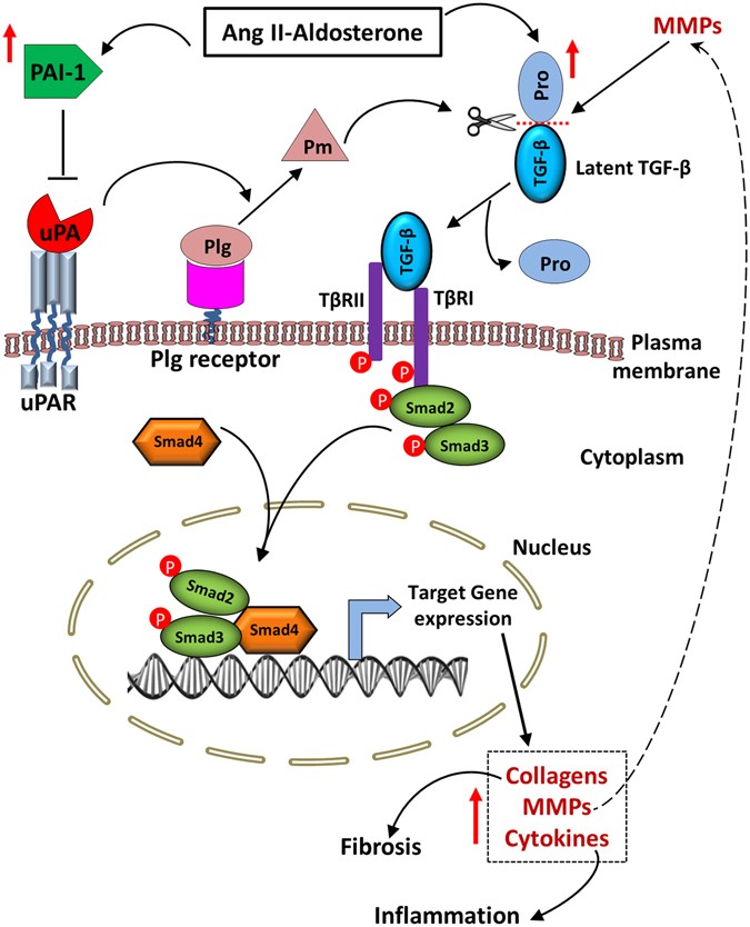 Schematic diagram showing functional roles of PAI-1 and uPA during AngII-Ald-induced cardiac fibrosis (Gupta et al., 2017)
Cancer:
Tumor Invasion: In certain cancer types, PAI-1 restrains tPA activity, limiting the capacity of cancer cells to invade neighboring tissues.
Tumor Microenvironment: PAI-1 plays a role in angiogenesis, facilitating the formation of new blood vessels that supply nutrients to tumors. It also fosters an immunosuppressive microenvironment that sustains tumor growth.
Metabolic Disorders:
Insulin Resistance: Elevated PAI-1 levels disrupt insulin signaling pathways, fostering insulin resistance and the onset of type 2 diabetes.
Obesity: PAI-1 is frequently overexpressed in adipose tissue among obese individuals, linking obesity to chronic inflammation and insulin resistance.
Liver Fibrosis:
Excessive Extracellular Matrix Accumulation: PAI-1 contributes to the undue accumulation of extracellular matrix proteins in the liver, leading to fibrosis. This can result from chronic liver damage caused by factors such as alcohol consumption or viral hepatitis.
Pulmonary Fibrosis:
Tissue Remodeling: In pulmonary fibrosis, PAI-1 participates in lung tissue remodeling. Elevated PAI-1 levels can result in the buildup of scar tissue, impairing lung function.
Obstructive Sleep Apnea (OSA):
Inflammation: OSA patients often exhibit heightened PAI-1 levels, which are associated with increased inflammation and oxidative stress, contributing to cardiovascular complications in these individuals.
Luminex Multiplex Technology for PAI-1 Signaling Pathway Analysis
The PAI-1 signaling pathway is a complex molecular network involving multiple proteins that interact and regulate each other. By employing Luminex multiplex technology, researchers can comprehensively examine the expression levels and post-translational modifications of key proteins in the PAI-1 pathway, shedding light on its intricate dynamics and functional relevance.
Experimental Workflow
Sample Preparation: The first step in the analysis involves preparing the biological samples of interest. These samples can include cell lysates, tissue extracts, or serum/plasma from patients with specific diseases or experimental treatments.
Bead-Based Assay: Luminex multiplex technology relies on microspheres or beads, each coated with specific capture antibodies targeting distinct proteins involved in the PAI-1 pathway. These beads are color-coded for identification.
Incubation: The prepared samples are then incubated with the bead mix containing the capture antibodies. During this step, proteins of interest in the samples bind to their respective capture antibodies on the beads.
Detection Antibodies: After incubation, biotinylated detection antibodies that recognize the same proteins are added. These antibodies create a sandwich complex with the target proteins, effectively labeling them.
Streptavidin-Phycoerythrin (SA-PE): Streptavidin-PE is introduced, binding to the biotinylated detection antibodies and forming a tripartite complex. This step further amplifies the signal.
Reading Fluorescence: The Luminex instrument detects the fluorescent signal from each bead, which corresponds to the amount of the target protein present in the sample. The color and fluorescence intensity of each bead identify the specific protein being measured.
Data Analysis: The generated fluorescence data are analyzed using specialized software. Standard curves, generated using known protein standards, are used to quantitate the protein levels in the samples. Data can be expressed in terms of concentration (pg/mL or ng/mL) or fold change relative to control samples.
Key Features
High Throughput Analysis: Luminex multiplex technology offers the advantage of high-throughput analysis, allowing researchers to assess multiple molecules within the PAI-1 pathway in a single experimental run. This not only saves time and resources but also enhances the efficiency of data collection.
Exceptional Sensitivity: The technology is highly sensitive, capable of detecting proteins at low concentrations. This sensitivity is crucial for studying signaling pathways like PAI-1, which involve molecules with varying expression levels.
Customization: Researchers have the flexibility to design customized panels of antibodies and probes tailored to their specific research questions. This customization enables the measurement of relevant proteins within the PAI-1 pathway, providing a targeted and informative analysis.
Multiplexing Capability: Luminex allows for the simultaneous analysis of multiple samples, making it ideal for comparative studies. This feature is particularly valuable when investigating differences in PAI-1 pathway activation across various experimental conditions or patient samples.
Reference:
- Gupta, Kamlesh K., et al. "Plasminogen activator inhibitor-1 protects mice against cardiac fibrosis by inhibiting urokinase-type plasminogen activator-mediated plasminogen activation." Scientific Reports 7.1 (2017): 365.