
Cell culture supernatant cytokine profiling is reliable only when collection time, culture exposure, cell-associated denominator, and media controls are planned together. A pg/mL value measures what accumulated in the collected medium; it does not necessarily measure cytokine secretion per viable cell or distinguish biological secretion from medium-associated background.
Multiplex profiling is particularly valuable when investigators need to read coordinated inflammatory, chemokine, and growth-factor responses from one conditioned-media sample. The results become difficult to interpret when groups differ in plating density, viability, growth rate, collection volume, or time in culture. Those variables should be recorded as part of the analytical design, not reconstructed later.
Key takeaways
- Choose sampling windows around expected secretion kinetics; do not assume one timepoint fits every cell system.
- Retain the measured pg/mL concentration, then add a prespecified cell-normalized result when cell number or viability can vary.
- Use matrix-matched media blanks and relevant unstimulated controls to separate secretion from medium background.
- When high- and low-abundance targets conflict, use pilot data to set dilution lanes or split panels before production.
Why Cell Culture Supernatant Cytokine Results Need More Than pg/mL
Concentration in supernatant depends on at least four linked factors: amount secreted, number of secreting cells, volume of medium, and collection interval. A lower concentration may represent reduced secretion, fewer viable cells, a larger collection volume, a shorter accumulation window, or a combination of these factors. Without a matching cell-associated measurement, the biological claim is often ambiguous.
Cell density is not merely a technical nuisance. In a macrophage model, cytokine secretion per stimulated cell changed with plating density even when the overall supernatant concentration appeared relatively stable (Vaughan-Jackson et al., 2022). This illustrates why two cultures with the same pg/mL output can have different cell-level secretion behavior.
For broad exploratory coverage from conditioned media, cell culture conditioned-media cytokine assays can be paired with a study-specific panel. The first planning question should be what the output is intended to represent: accumulated cytokine in a well, secretion per viable cell, or a time-resolved response curve.
How to Choose Cytokine Sampling Timepoints in Cell Culture
Cytokine release is kinetic. Some mediators rise quickly after stimulation and decline as they are consumed, degraded, or diluted by medium changes. Others accumulate over longer windows. A single terminal timepoint is useful only if it corresponds to the biological decision the study needs to make.
Match the timepoint to the intended claim
| Study question | Recommended timepoint logic | Key control |
|---|---|---|
| Early pathway activation | Use an early window informed by a small kinetic pilot. | Baseline and matched stimulation control. |
| Peak secretory response | Sample more than one interval to identify rise, peak, and decline. | Cell viability across the full window. |
| Accumulated output over exposure | Use one fixed, clearly defined collection interval. | Uniform medium volume and exposure duration. |
| Repeated sampling from one culture | Document every partial harvest and replacement volume. | Account for cumulative removal and dilution. |
A practical feasibility design often includes a limited number of representative conditions sampled at more than one timepoint. This is not a bench protocol; it is a risk-reduction step that reveals whether decision-critical cytokines are below range, saturated, or kinetically misaligned with the proposed collection window.
If the intended question concerns a response trajectory, select multiple biologically meaningful windows and analyse them as a time course. If the intended question concerns output after a standardized exposure, use a single fixed interval and keep it unchanged for every group. In either case, avoid interpreting a late accumulated pg/mL value as a direct measure of instantaneous secretion rate.
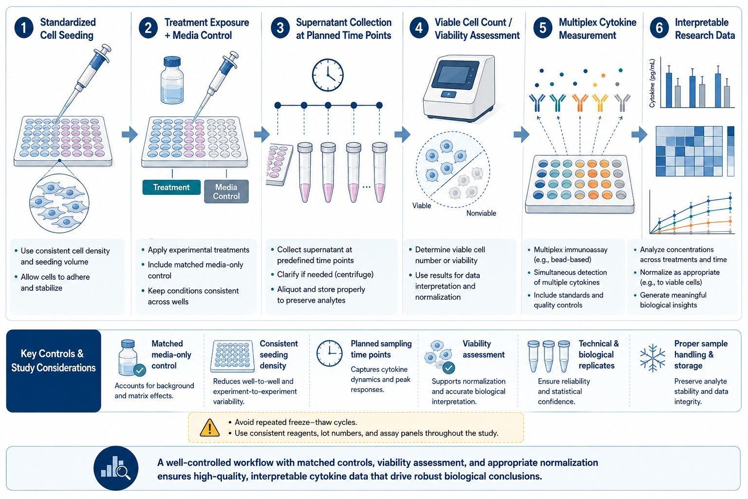 A conditioned-media cytokine study should align plating, exposure, collection time, cell-associated measurements, and reporting units.
Cell-Normalized Cytokine Results: Cell Count, Viability, and Protein Denominators
Keep the unadjusted concentration in pg/mL because it is the direct analytical result. Add a normalized result only when its denominator is measured consistently and answers a defined question. Common options include viable cell number at collection, starting plated cell number, total cell number, total DNA, or total cellular protein.
| Denominator | What it supports | Use with caution when |
|---|---|---|
| Endpoint viable cell number | Secretion relative to cells present at harvest; useful in cytotoxic or proliferative conditions. | Cell populations change composition or viability readout is not from the same unit. |
| Starting plated cell number | Standardized input across wells and early screening. | Large growth, death, detachment, or proliferation differences occur during exposure. |
| Total cell number | Useful where viability differences are minor and counting is consistent. | Dead cells or debris contribute substantially. |
| Total DNA or cellular protein | Alternative when direct cell counting is not practical. | Treatment changes DNA content, cell size, or protein content per cell. |
A clear reporting convention might provide: measured pg/mL, collection volume, collection interval, viable cells at endpoint, and calculated pg per 106 viable cells per interval. That presentation lets readers distinguish concentration in the medium from cell-associated output without concealing the raw result.
If expected treatments affect both cytokine secretion and survival, endpoint viable cell number should generally be recorded. If the study focuses on standardized cellular input with limited proliferation, starting cell number may be sufficient. If neither measure is available, report pg/mL transparently and limit the conclusion to concentration in the collected supernatant.
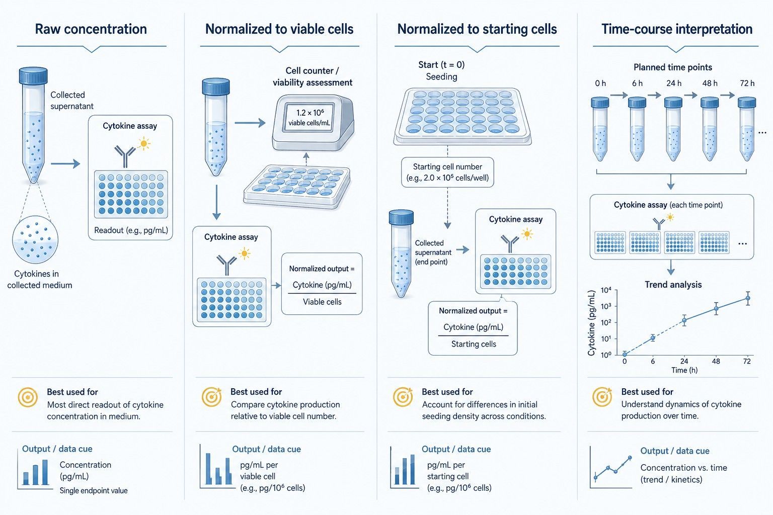 Raw concentration and a cell-associated denominator answer different questions and should be reported together when cell number can vary.
Supernatant Collection, Storage, and Batch Design
Collection handling should be standardized as tightly as the exposure condition. Record plate format, seeding density, medium volume, collection interval, whether samples were clarified, and the time from harvest to storage. These details determine whether an observed difference can be interpreted as a cellular response rather than a difference in sample handling.
When samples are collected over several days or plates, include matched controls on each collection batch. A media blank, unstimulated control, and a relevant responsive condition help distinguish a batch shift from a treatment effect. Keep the same medium formulation and supplement lot within a direct comparison when feasible; if that is not possible, treat lot or formulation as a documented design factor instead of an unrecorded nuisance.
Avoid deciding how to combine plates only after reviewing the cytokine results. Before collection, specify whether samples will be measured in one analytical run, in balanced batches, or with a bridge material repeated across batches. Include sample order and plate position in the data record. This becomes particularly important when a time course, several cell donors, or multiple treatment rounds must be compared.
For serial collections, distinguish an accumulated concentration from a new secretion interval. If medium is partially removed and replaced, record the removed and replacement volumes. A later sample then represents a new collection interval under altered culture conditions, not simply an extension of the first measurement. This distinction protects the interpretation of apparent increases or decreases over time.
Media Controls and Background Control for Multiplex Cytokine Assays
Conditioned medium contains more than secreted cytokines. Serum supplements, growth factors, albumin, phenol red, antibiotics, carrier proteins, and treatment formulations can contribute background or change assay behavior. A buffer-only blank is not always a sufficient control because it does not reproduce the full culture matrix.
Use controls that mirror the analytical question
- Media-only blank: the complete medium, including supplements, incubated and processed in parallel without cells.
- Unstimulated cell control: establishes cell-associated baseline output under the same culture conditions.
- Vehicle control: controls for formulation or solvent exposure where relevant.
- Cell-viability or count control: provides the denominator required to interpret secretion changes.
- Positive biological control: where scientifically justified, confirms that the culture system can generate a detectable response.
Background subtraction should not be automatic. First inspect whether the media blank is detectable for an analyte and whether it remains stable across runs. If a supplement-derived signal is present, describe the subtraction or reporting rule in advance and apply it consistently. If the background varies materially between media formulations, analyze those formulations separately rather than treating them as one matrix.
Multiplex and single-analyte measurements can show analyte-specific agreement rather than one universal level of interchangeability. For this reason, a culture-medium pilot should focus on the markers that drive the study decision and retain its dilution, blank, and control rules in the final sample manifest (duPont et al., 2005; Richens et al., 2010).
For questions where a treatment may create high cytokine output in some conditions and low output in others, consult the multiplex cytokine panel dilution and split strategy guide. That related resource addresses dynamic-range conflict; the present page addresses what the measured supernatant amount means biologically.
When to Use a Single Concentration, a Secretion Rate, or a Time-Course Readout
Choose one primary output before the study starts. A single pg/mL concentration works when plate density, volume, viability, and collection interval are tightly standardized and the question is accumulated mediator concentration. A cell-normalized amount works when groups may differ in cell number or viability. A time course works when the shape and timing of the response are part of the hypothesis.
If the study asks whether a stimulus changes total inflammatory milieu in a standardized well, choose fixed-window pg/mL with matched media controls. If it asks whether treatment changes cellular secretory capacity independent of cell loss, choose a viable-cell-normalized result alongside raw concentration. If it asks when an immune program rises or resolves, choose preplanned serial windows and do not collapse them into one terminal comparison.
Common Mistakes in Conditioned-Media Cytokine Studies
- Comparing pg/mL across wells with different cell numbers, viability, or collection volumes.
- Using a medium blank that lacks serum supplements or treatment vehicle present in the study samples.
- Selecting a single timepoint without confirming that critical cytokines are in a useful range.
- Normalizing only after a treatment appears cytotoxic or proliferative.
- Mixing samples collected over different exposure durations into one analysis.
- Interpreting decreased supernatant concentration as reduced secretion without a matched cellular readout.
Planning a Cell Culture Supernatant Cytokine Profiling Project
For scoping, provide cell type, species, culture medium and supplements, treatment conditions, planned collection windows, expected changes in viability or proliferation, sample number, available volume, and key cytokines. These inputs determine whether a broad multiplex panel, focused panel, one dilution, or a split design best protects the primary readouts.
Creative Proteomics can support custom cytokine panel design and conditioned-media profiling with fit-for-purpose collection, control, and normalization planning. The deliverable can retain raw pg/mL values, selected normalized outputs, media-control information, and QC annotations so the reported result matches the biological question.
Cell Culture Supernatant Cytokine Study Design Checklist
Before the production run, specify the culture plate format, seeded cell number, medium formulation and supplement lot, treatment and vehicle controls, collection volume, exact collection interval, clarification step, storage rule, media-only blank, and the cell-associated measurement that will support interpretation. In addition, identify which cytokines are decision-critical. The feasibility work should be designed around whether those markers remain within a useful measurement range, not around an average result across the whole panel.
For a clean final dataset, predefine how blank-associated values, measurements outside the reportable range, failed viability readouts, and repeated collections will be displayed. Retain the raw pg/mL result even when a normalized value is used for the primary comparison. This enables researchers to trace whether a normalized difference is driven by altered concentration, altered cell number, or both, and gives a service partner the information needed to recommend a focused follow-up experiment.
Frequently Asked Questions
Should cell culture supernatant cytokines be reported as pg/mL?
pg/mL is the measured concentration in the collected medium and should always be retained. When groups differ in cell number, viability, culture area, or collection volume, add a prespecified denominator such as pg per 10^6 viable cells or pg per well per defined collection interval.
How do I choose a cytokine sampling time?
Choose timepoints around the expected secretion kinetics and include an early feasibility panel rather than assuming one universal endpoint. If the question is peak response, sample the rise and decline; if the question is accumulated output, define a fixed collection window and document it consistently.
Why are media-only controls necessary for multiplex cytokine assays?
Media, serum supplements, growth factors, and additives can contribute background or interfere with measurement. A matrix-matched media blank processed alongside study samples helps distinguish biological secretion from medium-associated signal.
Can a lower cytokine concentration simply mean fewer cells survived?
Yes. A lower pg/mL value may reflect lower viable cell number, different confluence, or reduced culture duration rather than reduced secretion per cell. Pair supernatant data with a cell-count or viability readout collected on the same experimental unit.
Should I normalize to starting cell number or endpoint viable cell number?
The choice depends on the claim. Starting cell number supports standardization of input, while endpoint viable cell number better describes secretion relative to cells present at collection. If proliferation or toxicity is expected, record both and state which one governs the main analysis.
When should culture supernatant samples be diluted or split across panels?
Use a feasibility set when high-abundance factors may saturate while low-abundance factors remain near the lower range. If one dilution compromises decision-critical markers, split the panel or use separate dilution lanes rather than forcing all analytes into one condition.
References:
- Vaughan-Jackson A, et al. Density dependent regulation of inflammatory responses in macrophages. Frontiers in Immunology. 2022;13:895488.
- Cedrone E, et al. Multiplex enzyme-linked immunosorbent assay for detection of human cytokines in culture supernatants. National Cancer Institute Assay Cascade Protocols. 2020.
- DuPont NC, et al. Validation and comparison of Luminex multiplex cytokine analysis kits with ELISA in culture supernatants. Journal of Reproductive Immunology. 2005;66:175–191.
- Richens JL, et al. Quantitative validation and comparison of multiplex cytokine kits. Journal of Biomolecular Screening. 2010;15:562–568.
- Drewry AM, et al. Stimulated whole blood cytokine release: the need for a standardized methodology. Shock. 2016;45:490–499.